Search results (9 results)
-
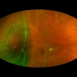 Rhegmatogenous Macula-On Retinal Detachment (Honeycomb)
Aug 6 2024 by Xitlali Caterina
Ultra-wide field fundus photograph of a 72 year old female with a macula-on retinal detachment with multiple breaks affecting her right eye. Patient presented in the office with flashes of light for five consecutive days prior. The patients vision was sc20/30 PHNI. The physician also noted an acute posterior vitreous detachment and lattice degeneration in the affect eye.
Photographer: Xitlali Caterina
Imaging device: Optos California RGB
Condition/keywords: honeycomb, lattice degeneration, Optos, posterior vitreous detachment, Retinal Detachment with Multiple Breaks, rhegmatogenous retinal detachment, ultra-wide field imaging
-
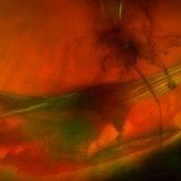 Vitreoretinal Traction with Adjacent Tear and Vitreous Hemorrhage
Oct 3 2023 by Alexis Singstock
Ultra-widefield fundus photograph of a 76 year old woman with vitreoretinal traction, an adjacent retinal tear and vitreous hemorrhage affecting the left eye. Patient was referred for retinal detachment and vitreous hemorrhage. Patient reports waking up the day prior to their appointment with "a lot of lines coming down the front, like swirling dirt in the left eye". Patient's vision was counting fingers at 1 ft. Dr. Joseph Boss noticed a horseshoe tear inferior to traction on exam and with the help of ultra-widefield imaging. Dr. Boss performed laser retinopexy to tear and impending tear at site of traction. Patient is scheduled for pars plana vitrectomy for dense vitreous hemorrhage.
Photographer: Alexis Singstock
Imaging device: Optos California
Condition/keywords: acute posterior vitreous detachment, fundus photography, left eye, Optos, OPTOS CALIFORNIA, pseudocolor, ULTRA WIDE FIELD, vitreoretinal traction, vitreous hemorrhage
-
 Inactive Toxoplasmosis
Nov 9 2012 by Norman Byer
This is the same case as in the previous photograph showing the very large free operculum torn from the retina.
Condition/keywords: acute posterior vitreous detachment, free operculum, inactive toxoplasmosis, tractional retinal tear
-
 Inactive Toxoplasmosis
Nov 9 2012 by Norman Byer
This 28-year-old man had inactive toxoplasmosis and presented with acute symptoms caused by this tractional retinal tear adjacent to a retinochorodial scar. He also had an acute posterior vitreous detachment which had torn this retinal operculum completely free. The next slide shows the same lesion. Note the early rolled edge on the left side of the tear.
Condition/keywords: acute posterior vitreous detachment, inactive toxoplasmosis, operculum, rolled edges of retina, tractional retinal tear
-
 Acute Posterior Vitreous Detachment
Nov 9 2012 by Norman Byer
This large and complicated retinal tear in a 51-year-old man resulted from an acute posterior vitreous detachment which concentrated its tractional forces around this area of lattice degeneration. Because of the powerful traction, there is an additional central tear splitting the large retinal flap and almost severing one of its arms. The traction was strong enough to completely rupture the blood vessel just to the left of the flap. Marking the ruptured peripheral end of the blood vessel is a yellow depigmented thrombus.
Condition/keywords: acute posterior vitreous detachment, depigmented thrombus, lattice degeneration, retinal tear, tractional retinal detachment
-
 Posterior Vitreous Detachment
Nov 9 2012 by Norman Byer
In this 50-year-old man, these two adjacent tears are separated by a narrow band of tissue but have a common flap. They were caused by a posterior vitreous detachment and they are surrounded by a localized area of detachment. This case is similar to slide pair 53.
Condition/keywords: acute posterior vitreous detachment, adjacent tears, bridge of tissue between tears, posterior vitreous detachment, retinal tear
-
 Vitreous Hemorrhage
Nov 9 2012 by Norman Byer
This 60-year-old man suddenly developed a vitreous hemorrhage from this acute horseshoe tear 3½ years following cataract extraction when a posterior vitreous detachment occurred. The white nubbin identifies this lesion as a preexisting cystic retinal tuft. The pigment spot beneath the flap is evidence of secondary trophic changes in the pigment epithelium. Note the irregular shape of the flap with the narrow tip and broad base. This was caused by vitreous traction which was exerted at two separate points on the retina and which tore the retina at each place.
Condition/keywords: acute posterior vitreous detachment, irregularly shaped flap, trophic pigmented changes, vitreous hemorrhage, vitreous traction, white retinal tuft
-
 Symptomatic Retinal Tear
Nov 9 2012 by Norman Byer
This is another example of a symptomatic retinal tear which occurred at the site of a cystic retinal tuft two days prior to the photograph when an acute posterior vitreous detachment occurred in this 64-year-old woman. Note the horizontal line of vitreous blood along the lower edge of the flap which demarcates the vitreous attachment to the flap.
Condition/keywords: acute posterior vitreous detachment, cystic retinal tuft, retinal flap, retinal tear, vitreous blood
-
 Cystic Retinal Tuft
Nov 9 2012 by Norman Byer
This is the same lesion as in the previous slide pair but the photograph was taken nine years later when the patient was 58-years-old soon after an acute posterior vitreous detachment. This demonstrates that posterior vitreous detachment can produce large retinal tears at these sites. However, it is important to emphasize that prophylactic treatment of cystic retinal tufts in the absence of a retinal tear would be very ill-advised because several hundred innocence and harmless lesions would have to be treated in order to prevent one tear of the retina.
Condition/keywords: cystic retinal tuft, posterior vitreous detachment, retinal tear
 Loading…