Search results (402 results)
-
 Not All Stars Are in the Sky — Some Live in the Eyes of Those Learning to See in New Ways
Apr 21 2025 by rohan jain
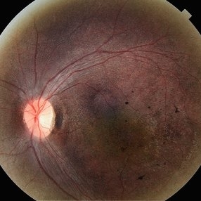
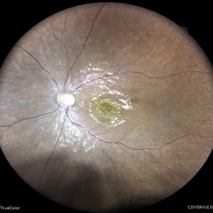
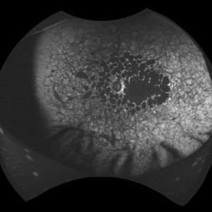
Stargardt disease
Photographer: Dr. ROHAN JAIN
Condition/keywords: fleck retinopathy, fundus autofluorescence (FAF), hereditary macular dystrophy
-
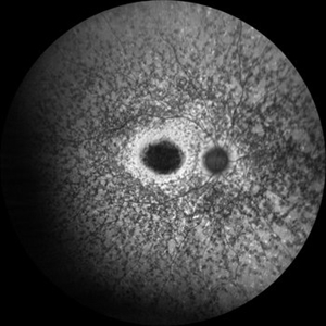
 Stargardt Disease (FA)
Jan 22 2025 by Virginia Gebhart
Fluorescein angiogram of 19 year old female with confirmed Stargardt Disease. Hyperfluorescence in the macula with staining defect and silent choroid.
Photographer: Virginia Gebhart, Retina Consultants of Carolina
Imaging device: Optos California
Condition/keywords: fluorescein angiogram (FA), Silent Choroid, Stargardt disease
-
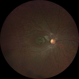
 Stargardt Disease
Jan 22 2025 by Virginia Gebhart
19 year old female with confirmed Stargardt Disease. Central RP atrophy with pigment clumping and "beaten metal" appearance. BCVA 20/125
Photographer: Virginia Gebhart, Retina Consultants of Carolina
Imaging device: Topcon 50DX
Condition/keywords: pigment clumps, RP atrophy, Stargardt disease
-
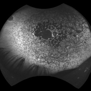
 Stargardt's Disease
Oct 23 2024 by Virginia Gebhart
62 year old female with bullseye RPE changes and flecks, mottled FAF, and silent choroid on FA consistent with late onset Stargardt's Disease. Pt is asymptomatic with 20/20 vision OU at this time
Photographer: Virginia Gebhart, Retina Consultants of Carolina
Imaging device: Optos California
Condition/keywords: Stargardt disease, Stargardts Disease
-
 Silent Choroid
Oct 10 2024 by Philip Conkling, MD
Fluorescein angiogram demonstrating silent choroid in Stargardt disease.
Imaging device: Optos
Condition/keywords: Stargardt disease
-
 Silent Choroid
Oct 10 2024 by Philip Conkling, MD
Fluorescein angiogram demonstrating silent choroid in Stargardt disease.
Imaging device: Optos
Condition/keywords: Stargardt Disease
-
 Stargardt Disease
Oct 10 2024 by Philip Conkling, MD
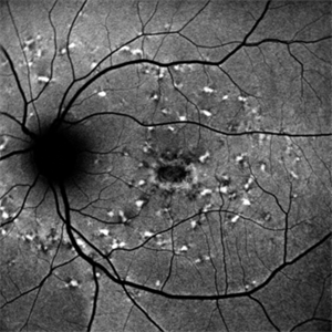
Fundus autofluorescence demonstrating typical findings of stargardt disease.
Imaging device: Optos
Condition/keywords: Stargardts Disease
-
 Stargardt Disease
Oct 10 2024 by Philip Conkling, MD
Fundus autofluorescence demonstrating typical findings of stargardt disease.
Imaging device: Optos
Condition/keywords: Stargardt Disease
-
 Pattern dystrophies – Asymptomatic middle-aged man with normal vision and a multifocal PD
Sep 17 2024 by Nicolas A Yannuzzi, MD
The PD simulates Stargardt disease/fundus flavimaculatus with irregular yellow-white flecks scattered throughout the posterior pole. Some lesions extend beyond the retinal vascular arcades.
Condition/keywords: inherited retinal disease, pattern dystrophy
-
 Pattern dystrophies – OCT demonstrates significant RPE irregularities and multiple focal inner segment-outer segment (IS-OS) disruptions with overlying cystic changes in both eyes of a 60-year-old man with PD
Sep 17 2024 by Nicolas A Yannuzzi, MD
Visual acuity was 20/20 OD and 20/25 OS. Genetic testing showed only 1 pathogenic variation in ABCA4, which is atypical for STGD1 Stargardt disease that is inherited in autosomal recessive fashion. (Images courtesy of Byron L. Lam, MD)
Condition/keywords: inherited retinal disease, pattern dystrophy
-
 Stargardt Disease
Aug 29 2024 by César Adrián Gómez Valdivia, MD
Fundus photograph of a 10 year-old male patient with Stargardt Disease. Findings were Bilateral.
Photographer: @eyemissu2
Imaging device: TOPCON TRC-50DX
Condition/keywords: Stargardt disease
-
 Stargardt Disease
Aug 27 2024 by Korey Starkey
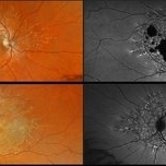
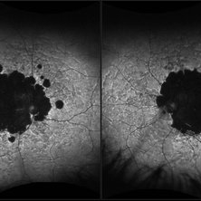
Ultra wide-field fundus photograph and fundus autofluorescence of a 49-year-old male. Initial visit imaging.
Photographer: Korey Starkey
Imaging device: Optos
Condition/keywords: fundus autofluorescence (FAF), fundus photograph, Optos, Stargardt disease, ultra-wide field imaging
-
 B-FAF in Stargardt's Disease
Jul 4 2024 by Tejaswita Verma
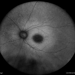
Blue fundus autofluorescence showing hypoautofluorescence picture of a 28 year old male with 6/60 vision in BE in a case of Stargardt's disease.
Photographer: DR. TEJASWITA VERMA
Imaging device: MIRANTE
Condition/keywords: fundus autofluorescence (FAF), hereditary macular dystrophy, Stargardt disease
-
 Representative Multifocal Electroretinography Responses
May 13 2024 by Gabrielle Hallai
Multifocal ERG responses from a control individual with no known retinal pathology is shown on the left. The topographical maps (left of each panel) demonstrate the patient’s pattern of responses. The trace arrays (right of each panel) demonstrate the patient’s multifocal ERG responses. The middle set of images demonstrates responses from a patient with Stargardt disease. The topographical map shows decreased patterns throughout the macula. The traces show decreased central response with preserved, but diminished responses in the periphery. The final set of images is from a patient with retinitis pigmentosa. In this case, the topographical map shows a small, green peak in the center. In the trace array, there are extinguished responses in the periphery with a diminished response in the center. Multifocal ERG testing was completed using the Diagnosys LCD Pattern Stimulator.
Photographer: Gabrielle Hallai, PhD, Cleveland Clinic Cole Eye Institute
Imaging device: Diagnosys LCD Pattern Stimulator
Condition/keywords: electroretinography, multifocal ERG (MFERG), retinitis pigmentosa, Stargardt disease
-
 Stargardt's Disease
Apr 20 2024 by Tejaswita Verma
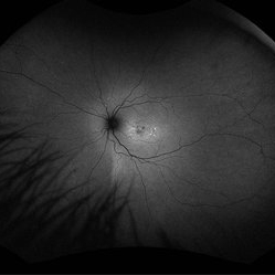
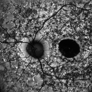
Fundus autofluorescence image of the left eye of a 39 year old male showing hypoautofluorescence in a case of Stargardt's disease.
Photographer: DR. TEJASWITA VERMA
Imaging device: MIRANTE
Condition/keywords: fundus autofluorescence (FAF), hereditary macular dystrophy, hypoautofluorescence, Stargardt disease
-
 Stargardt's Disease
Apr 20 2024 by Tejaswita Verma
Fundus autofluorescence image of the right eye of a 39 year old male showing hypoautofluorescence in a case of Stargardt's disease.
Photographer: DR. TEJASWITA VERMA
Imaging device: MIRANTE
Condition/keywords: fundus autofluorescence (FAF), hereditary macular dystrophy, hypoautofluorescence, Stargardt disease
-
 Stargardts' Disease
Apr 9 2024 by Akansha Sharma
Autofluorescence image of a 28 year old male with stargardts' disease.
Photographer: Dr. Akansha Sharma, Bharati Eye Hospital
Condition/keywords: Stargardt disease
-
 Stargardts' Disease
Apr 9 2024 by Akansha Sharma
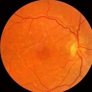
Color fundus photograph of a 28 year old male with stargardts' disease.
Photographer: Dr. Akansha Sharma, Bharati Eye Hospital
Condition/keywords: Stargardt disease
-
 Stargardt Disease
Apr 8 2024 by T. P . VIGNESH, MBBS,MS
Fundus autofluorescence image of the left eye revealing foveal hypoautofluorescence and multiple hypoautofluorescent specks in the background radiating from posterior pole towards periphery .
Photographer: Bharathi
Imaging device: ZEISS CLARUS
Condition/keywords: fundus autofluorescence (FAF), Stargardts Disease
-
 Stargardt Disease
Apr 8 2024 by T. P . VIGNESH, MBBS,MS
Fundus autofluorescence image of the right eye revealing foveal hypoautofluorescence and multiple hypoautofluorescent specks in the background radiating from posterior pole towards periphery.
Photographer: Bharathi
Imaging device: ZEISS CLARUS
Condition/keywords: fundus autofluorescence (FAF), Stargardt disease
-
 Stargardt Disease
Apr 8 2024 by T. P . VIGNESH, MBBS,MS
Fundus photo of left eye of 20 year old woman revealing beaten bronze foveal atrophy and wide-spread flecks radiating from the posterior pole to the periphery .
Photographer: Bharathi
Imaging device: ZEISS CLARUS
Condition/keywords: fundus flavimaculatus, Stargardt disease, Stargardts Disease
-
 Stargardt Disease
Apr 8 2024 by T. P . VIGNESH, MBBS,MS
Fundus photo of right eye of 20 year old woman revealing beaten bronze foveal atrophy and wide-spread flecks radiating from the posterior pole to the periphery .
Photographer: Bharathi
Imaging device: ZEISS CLARUS
Condition/keywords: Stargardt disease
-
 Stargardt Disease
Apr 2 2024 by José Laércio Araújo Filho
Fundus autofluorescence of a 43-year-old woman with a ABCA4 positive macular dystrophy compatible with Stargardt Disease.
Photographer: José Laércio de Araújo Filho, Universidade de São Paulo, São Paulo
Imaging device: Optos Daytona P200T / A10600
Condition/keywords: cone dystrophy, Stargardt disease
-
 Advanced Stargardt Disease
Mar 25 2024 by Angela Rico
69 yr old female who presents with VA: OD 20/CF@4', OS 20/250.
Photographer: Angela Rico M.D., Retina Specialists of Tampa
Condition/keywords: Stargardts Disease
 Loading…