Search results (26 results)
-
 Unifocal RPE Hypertrophy
Apr 2 2019 by Gary R. Cook, MD, FACS
40-year-old white female with a well-demarcated area of unifocal RPE hypertrophy lacking the typical vacuoles seen in CHRPE lesions superiorly; V.A. = 20/15
Imaging device: Topcon VT-50
Condition/keywords: congenital hypertrophy of the retinal pigment epithelium (CHRPE), hypertrophy
-
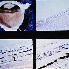 Slide 9-60
Feb 26 2019 by Lancaster Course in Ophthalmology
Diffuse peripheral RPE hypertrophy. There is a band of pigmentation just posterior to the ora serrata (upper left) where the RPE is darker and contains larger, spherical pigment granules(lower right). The junction (arrow) between normal (left) and hypertrophic (right) pigment epithelium is illustrated in the lower left view. A few areas of paving-stone degeneration are present at the equator (upper left).
Condition/keywords: ora serrata, retinal pigment epithelium (RPE) hypertrophy
-
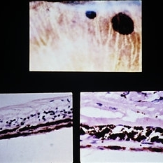 Slide 9-59
Feb 26 2019 by Lancaster Course in Ophthalmology
Two localized areas of RPE hypertrophy. The RPE has melanin granules that are large and spherical (lower views.)
Condition/keywords: melanin granules, retinal pigment epithelium (RPE) hypertrophy
-
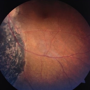
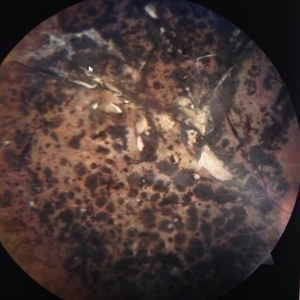
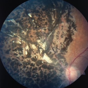
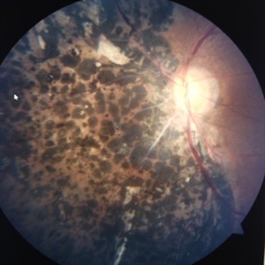
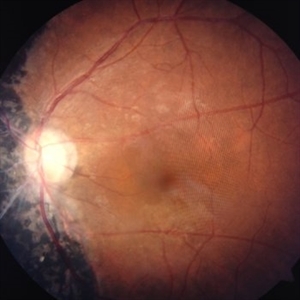
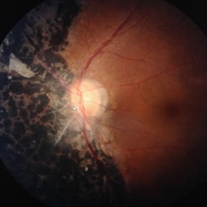
 Self-Applied Retinal Detachment
Sep 24 2017 by Ivonne Jocelyn Rivera Alvarado
40-year-old female, asymptomatic, without history of trauma. VA 20/20. No comorbilities. No ophthalmologic surgeries. It was a incidental finding. It can be observed a large RPE hypertrophy at the nasal retinal zone that borders the optic nerve with a line of demarcation that corresponds to a self applied retinal detachment.
Photographer: Ivonne Jocelyn Rivera Alvarado, Tec de Monterrey, Mexico
Condition/keywords: retinal pigment epithelium (RPE) hypertrophy
-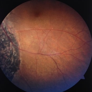
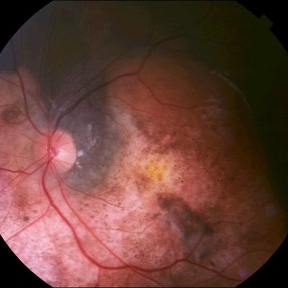 Peripapillary Choroidal Osteoma
Oct 2 2015 by Paul T. Finger, MD, FACS
Note the lightly albeit variably pigmented peripapillary tumor. It is relatively flat, with overlying evidence of RPE hypertrophy. Note the scalloped edges. CNV is not seen in this case.
Photographer: anonymous
Condition/keywords: choroidal osteoma
-
 PE Hypertrophy
Oct 6 2014 by Howard Schatz, MD
55-year-old white female. PE Hypertrophy. Re 20/13 LE 20/16.
Condition/keywords: PE hypertrophy
-
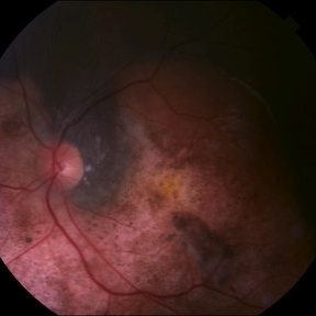 Choroidal Osteoma
Aug 29 2014 by Paul T. Finger, MD, FACS
Note the relatively flat, yellow-white tumor with overlying clusters of RPE hypertrophy and scalloped edges. This choroidal osteoma also had CNV that responded to photodynamic therapy.
Imaging device: Topcon
Condition/keywords: choroidal osteoma
-
---thumb.jpg/image-square;max$300,300.ImageHandler) RPE Hypertrophy
RPE Hypertrophy
Aug 8 2013 by From the Collections of Thomas M. Aaberg, MD and Thomas M. Aaberg Jr., MD
unknown
Condition/keywords: retinal pigment epithelium (RPE) hypertrophy
-
---thumb.jpg/image-square;max$300,300.ImageHandler) RPE Hypertrophy In The Posterior Pole
RPE Hypertrophy In The Posterior Pole
Aug 8 2013 by From the Collections of Thomas M. Aaberg, MD and Thomas M. Aaberg Jr., MD
Multiple congenital RPE hypertrophy with more prominent atrophic component (lacuna) in the macula and inferior arcade.
Condition/keywords: retinal pigment epithelium (RPE) hypertrophy
-
---thumb.jpg/image-square;max$300,300.ImageHandler) Congenital RPE Hypertrophy
Congenital RPE Hypertrophy
Aug 8 2013 by From the Collections of Thomas M. Aaberg, MD and Thomas M. Aaberg Jr., MD
unknown
Condition/keywords: retinal pigment epithelium (RPE) hypertrophy
-
---thumb.jpg/image-square;max$300,300.ImageHandler) Congenital RPE Hypertrophy
Congenital RPE Hypertrophy
Aug 8 2013 by From the Collections of Thomas M. Aaberg, MD and Thomas M. Aaberg Jr., MD
Well - demarcated CHRPE inferonasal to the optic disc of right eye.
Condition/keywords: retinal pigment epithelium (RPE) hypertrophy
-
---thumb.jpg/image-square;max$300,300.ImageHandler) RPE Hypertrophy
RPE Hypertrophy
Aug 8 2013 by From the Collections of Thomas M. Aaberg, MD and Thomas M. Aaberg Jr., MD
FA of the same patient. Shows typical window defects in the lacunar area due to chorioretinal atrophy #2.
Condition/keywords: retinal pigment epithelium (RPE) hypertrophy
-
---thumb.jpg/image-square;max$300,300.ImageHandler) Congenital RPE Hypertrophy
Congenital RPE Hypertrophy
Aug 8 2013 by From the Collections of Thomas M. Aaberg, MD and Thomas M. Aaberg Jr., MD
Well demarcated congenital RPE hypertrophy with typical halo surrounding lesion and lacuna inside the lesion #1.
Condition/keywords: retinal pigment epithelium (RPE) hypertrophy
-
---thumb.jpg/image-square;max$300,300.ImageHandler) RPE Hypertrophy
RPE Hypertrophy
Aug 8 2013 by From the Collections of Thomas M. Aaberg, MD and Thomas M. Aaberg Jr., MD
Pathology slide shows RPE hypertrophy. (Melanin granules visible inside the RPE cells)
Condition/keywords: retinal pigment epithelium (RPE) hypertrophy
-
---thumb.jpg/image-square;max$300,300.ImageHandler) RPE Hypertrophy
RPE Hypertrophy
Aug 8 2013 by From the Collections of Thomas M. Aaberg, MD and Thomas M. Aaberg Jr., MD
Well demarcated round congenital hypertrophy of RPE with a narrow halo of hypopigmentation.
Condition/keywords: retinal pigment epithelium (RPE) hypertrophy
-
---thumb.jpg/image-square;max$300,300.ImageHandler) RPE Hypertrophy
RPE Hypertrophy
Aug 8 2013 by From the Collections of Thomas M. Aaberg, MD and Thomas M. Aaberg Jr., MD
Typical bear tracks in RPE hypertrophy.
Condition/keywords: retinal pigment epithelium (RPE) hypertrophy
 Loading…