Search results (116 results)
-
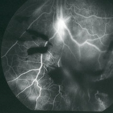
 Eales Disease
Jan 31 2025 by Thirumalesh Mochi Basavaraj, MD
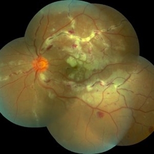
Ultra wide field image of a 24 year-old young healthy adult male with a visible sea fan neovascularization with partial PVD secondary to Scatter LASER photocoagulation with Vitreous and subhyaloid hemorrhage.
Photographer: Puttaswamy N K
Condition/keywords: Eales disease, Neovascularisation elsewhere (NVE), sea fan
-
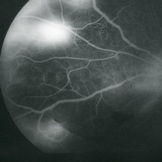
 Eales Disease
Jan 31 2025 by Thirumalesh Mochi Basavaraj, MD
Ultra-wide field image of a 24 year old young healthy adult male with a visible sea fan neovascularization with partial PVD with vitreous and subhyaloid hemorrhage.
Photographer: Puttaswamy
Condition/keywords: Eales disease, sea fan, Ultra-wide field retinal imaging
-
 Idiopathic Retinal Vasculitis
Jun 9 2024 by Anjana Mirajkar, MS Ophthalmology
A widefield image of a 32 year old male of LE showing sclerosed vessels more prominent inferiorly with superficial hemorrhages noted in all quadrants along with sheathing of vessels noted in superiorly.
Photographer: Dr. Anjana Mirajkar -Retina Foundation, Ahmedabad
Imaging device: Mirante-Nidek
Condition/keywords: Eales disease
-
Tractional RD-Making the Decision When and Where to Stop
May 23 2024 by ARVIND JAIN M
This is a young gentlemen with defective vision for 3 months in his right eye. He gave the history of recurrent redness of the right past few months. he was diagnosed to have right eye vasculitis with tractional detachment. He underwent uveitic workup and under steroid cover right eye paraplana vitrectomy with membrane peeling with endolaser with c3f8 gas was planned. patient improved significantly. this surgical video demonstrates when and where to stop during membrane peeling and get good results.
Condition/keywords: Eales disease, retinal vasculitis, tractional retinal detachment
-
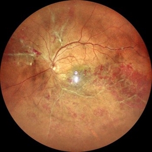
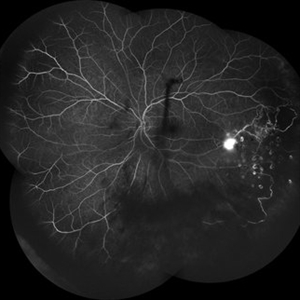
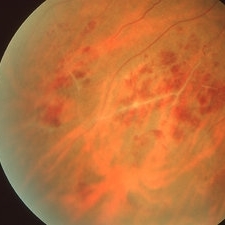
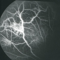
 Eales Disease
Feb 13 2024 by Narciso F. Atienza, MD, MBA, FASRS, FPCS, FPAO.
Angiographic study of a 31 year old male patient who came in with blurring of vision of the right eye. Colored photo shows neovascularization emanating from the optic nerve with scattered retinal hemorrhages distal to the noevascularization on the infero-nasal area, and temporal area. Areas of perivascular sheathing are also noted on the infero-temporal area and macula. Angiography shows neovascularization and areas of non-perfusion on the infero-nasal and peripheral temporal retina.
Photographer: Narciso F. Atienza, Jr, MD MBA, Legazpi Eye Center
Imaging device: Zeiss Clarus
Condition/keywords: Eales disease
-
 Eales Disease
Feb 13 2024 by Narciso F. Atienza, MD, MBA, FASRS, FPCS, FPAO.
Angiographic study of a 31 year old male patient who came in with blurring of vision of the right eye. Colored photo shows neovascularization emanating from the optic nerve with scattered retinal hemorrhages distal to the noevascularization on the infero-nasal area, and temporal area. Areas of perivascular sheathing are also noted on the infero-temporal area and macula. Angiography shows neovascularization and areas of non-perfusion on the infero-nasal and peripheral temporal retina
Photographer: Narciso F. Atienza, Jr, MD MBA, Legazpi Eye Center
Imaging device: Zeiss Clarus
Condition/keywords: Eales disease
-
 Eales' disease
Dec 12 2022 by Pramod Kumar Suman, MBBS, MD
Montage photograph wide field angiogram of an 32-year-old male with blocked fluorescence with leakage with peripheral capillary non perfusion areas.
Photographer: Pramod Kumar Suman, Retina Foundation, Ahmedabad
Imaging device: Mirante
Condition/keywords: Eales disease
-
 Eales' disease
Dec 12 2022 by Pramod Kumar Suman, MBBS, MD
Fundus photograph of an 32-year-old male with inferior vitreous hemorrhage.
Photographer: Pramod Kumar Suman, Retina Foundation, Ahmedabad
Imaging device: Mirante
Condition/keywords: Eales disease
-
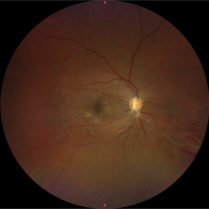
 Eales Disease
May 23 2021 by Katia Delalibera Pacheco, MD
Color fundus photograph of the left eye of a 37-year-old man with Eales disease. Note the peripheral to mid-peripheral periphlebitis in multiple quadrants concurrently. Venous dilation and perivascular exudate can be observed. We can also note the demarcation between perfused and nonperfused retina.
Photographer: CBV- Eye Hospital, Brasilia, DF, Brazil
Condition/keywords: Eales disease, occlusive retinal vasculitis
-
 Eales Disease
May 23 2021 by Katia Delalibera Pacheco, MD
Color fundus photograph of the left eye of a 37-year-old man with Eales disease. Note the peripheral to mid-peripheral periphlebitis in multiple quadrants concurrently. Venous dilation and perivascular exudate can be observed. We can also note the demarcation between perfused and nonperfused retina.
Photographer: CBV- Eye Hospital Brasilia, DF, Brazil
Condition/keywords: Eales disease
-
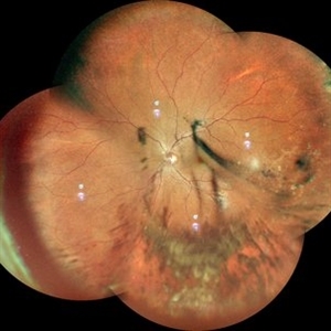
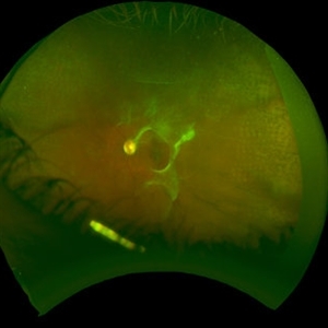 Eales Disease Causing TRD and Macular Edema in Pregnancy
Apr 21 2020 by Richard M Martindale, MD
42-year-old pregnant African American with TRD and peripheral ischemia secondary to Eales disease. She was assigned this diagnosis of exclusion after a thorough work up for other identifiable causes of peripheral ischemia (e.g. sickle cell, syphilis, sarcoid, clotting disorders, SLE, TB, IP, FEVR). We elected to temporize her with PRP and Ozurdex in lieu of anti-VEGF medication given her pregnant status. Note: the Ozurdex pellet is visible in the inferior aspect of this photo.
Photographer: Retina Consultants of Alabama
Imaging device: Optos
Condition/keywords: Eales disease
-
 Eales Disease
Apr 3 2019 by Paola Brito, MD
8-year-old girl with positive Matoux test. She received laser in nasal retina. Peripheral vein occlusion, ischemic areas and neovascularization.
Photographer: Paola Brito, Hospital de la Luz, Mexico
Imaging device: retcam
Condition/keywords: Eales disease
-
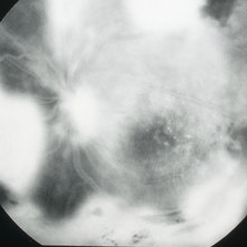 Eales Disease
Apr 1 2019 by Gary R. Cook, MD, FACS
Late-phase fluorescein angiogram image of the left eye of a 23-year-old Vietnamese female with Eales Disease showing extensive dye leakage from multiple areas of NVE and from some NVD.
Imaging device: Topcon VT-50
Condition/keywords: Eales disease, FA late phase, fluorescein angiogram (FA), neovascularization elsewhere (NVE)
-
 Eales Disease
Apr 1 2019 by Gary R. Cook, MD, FACS
Mid-phase fluorescein angiogram frame of the left eye of a 23-year-old Vietnamese female with Eales Disease showing multiple areas of NVE and areas of capillary loss and nonperfusion OS.
Imaging device: Topcon VT-50
Condition/keywords: Eales disease, FA mid phase, fluorescein angiogram (FA), neovascularization elsewhere (NVE)
-
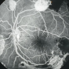
 Eales Disease
Apr 1 2019 by Gary R. Cook, MD, FACS
25-year-old Vietnamese male with peripheral retinal vasculitis, vaso-occlusion, and retinal hemorrhages secondary to Eales Disease; V.A.= 20/20.
Imaging device: Topcon VT-50
Condition/keywords: Eales disease, retinal hemorrhage, vaso-occlusive disease, vasoocclusive retinopathy
-
 Eales Disease
Apr 1 2019 by Gary R. Cook, MD, FACS
Mid-phase fluorescein angiogram image of the left eye of a 23-year-old Vietnamese female with Eales Disease showing the retinal vascular abnormalities, capillary loss, and a focus of NVE; V.A.= 20/25-2.
Imaging device: Topcon VT-50
Condition/keywords: Eales disease, FA mid phase, fluorescein angiogram (FA), neovascularization elsewhere (NVE)
-
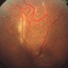
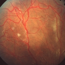 Eales Disease
Apr 1 2019 by Gary R. Cook, MD, FACS
Fundus photograph of the superonasal mid-periphery of a 20-year-old Vietnamese male with Eales Disease; V.A.= 20/40-1.
Imaging device: Topcon VT-50
Condition/keywords: Eales disease
-
 Eales Disease
Apr 1 2019 by Gary R. Cook, MD, FACS
Late-phase (5 minutes) fluorescein angiogram image of a 20-year-old Vietnamese male with Eales Disease showing retinal vascular changes and intense leakage from peripheral NVE.
Imaging device: Topcon VT-50
Condition/keywords: Eales disease, FA late phase leakage, fluorescein angiogram (FA), neovascularization elsewhere (NVE)
-
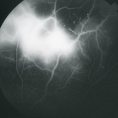
 Eales Disease
Apr 1 2019 by Gary R. Cook, MD, FACS
Mid-phase (70 seconds) fluorescein angiogram image of the inferior periphery OS of a 20-year-old Vietnamese male with Eales Disease; there is bright hyperfluorescence from a focus of NVE below the optic disc and blocked fluorescence from vitreous hemorrhage in the eye.
Imaging device: Topcon VT-50
Condition/keywords: Eales disease, FA late phase, FA late phase leakage, fluorescein angiogram (FA), neovascularization elsewhere (NVE), vitreous hemorrhage
-
 Eales Disease
Apr 1 2019 by Gary R. Cook, MD, FACS
Fundus photograph showing the retinal vascular abnormalities in the superior mid-periphery of a 20-year-old Vietnamese male with Eales Disease; V.A. = 20/40-1.
Imaging device: Topcon VT-50
Condition/keywords: Eales disease
-
 Eales Disease
Apr 1 2019 by Gary R. Cook, MD, FACS
Late-phase (5 minutes) fluorescein angiogram image of the nasal mid-periphery of the left eye of a 23-year-old Vietnamese female with Eales Disease showing multiple areas of NVE and some disc leakage.
Imaging device: Topcon VT-50
Condition/keywords: Eales disease, FA late phase, FA late phase leakage, fluorescein angiogram (FA), neovascularization elsewhere (NVE)
 Loading…