Search results (120 results)
-
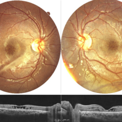
 Choroidal Melanoma with Exudative Detachment
Apr 7 2025 by Virginia Gebhart
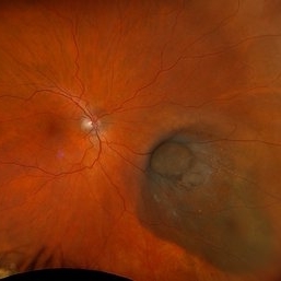
Autofluorescence image of 36 year old female showing demarcation line of fluid/detachment from new choroidal melanoma. Pt will be scheduled for brachytherapy pending CT scan results.
Photographer: Virginia Gebhart, Retina Consultants of Carolina
Imaging device: Optos California
Condition/keywords: Autoflourescence, autofluorescence imaging, choroidal melanoma, melanoma, retinal detachment
-
 New Choroidal Melanoma with Exudative Detachment
Apr 7 2025 by Virginia Gebhart
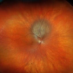
36 year old female referred for pigmented mass. Pt complains of flashes of light since last fall. Clinical exam and ultrasound findings consistent with choroidal melanoma with exudative detachment inferior. Pt will be scheduled for brachytherapy and possible tumor biopsy pending CT scan results.
Photographer: Virginia Gebhart, Retina Consultants of Carolina
Imaging device: Optos California
Condition/keywords: Choroidal melanoma, exudative detachment, melanoma, retinal detachment
-
 Solar Retinopathy
Apr 1 2025 by Isaac Agranoff
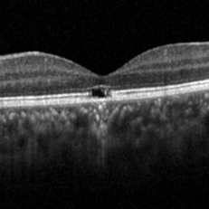
OCT scan of 18-year-old male presenting with 20/40 BCVA OU and bilateral focal outer retinal subfoveal defects. Patient reported long-term history of frequent sungazing, has stopped within past 6-9 months.
Photographer: Isaac Agranoff
Imaging device: Heidelberg Spectralis
Condition/keywords: solar retinopathy
-
 Collar Button Melanoma
Mar 27 2025 by Virginia Gebhart
62 year old male with large pigmented lesion with collar button. Pt states he was never aware of any lesion/nevus in the past. Fluid and orange pigment present, appears to be chronic. Pt will be scheduled for brachytherapy pending CT scan results.
Photographer: Virginia Gebhart, Retina Consultants of Carolina
Imaging device: Optos California
Condition/keywords: choroidal melanoma, collar button
-
 Ciliary Body Metastasis
Mar 26 2025 by Virginia Gebhart
54 year old female referred for iris mass. UBM shows large solid mass originating in the ciliary body and eroding into the anterior chamber under the iris epithelium. Recent CT scans revealed multiple bilateral pulmonary and hepatic nodules. Pt has been scheduled for PET scan and liver biopsy by radiation oncologist.
Photographer: Virginia Gebhart, Retina Consultants of Carolina
Imaging device: Samsung Galaxy
Condition/keywords: choroidal metastasis, ciliary body mass, metastatic cancer
-
 Choroidal Melanoma
Mar 10 2025 by Virginia Gebhart
56 year old female with new choroidal melanoma. Pt states they have a "freckle" that had been monitored for 26 years, last CEE was over 2 years ago. Clinical exam and ancillary testing consistent with uveal melanoma. Pt scheduled for plaque brachytherapy with transretinal biopsy of the tumor for genetic testing. Pt also scheduled for CT scan of chest/abdomen to rule out metastatic disease.
Photographer: Virginia Gebhart, Retina Consultants of Carolina
Imaging device: Optos California
-
 Ciliary Body Melanoma
Feb 12 2025 by Virginia Gebhart
91 year old female with large collar button tumor emanating from the ciliary body with resolving vitreous hemorrhage. Melanoma cells in the AV as well as studded on the entire retina surface. Pt scheduled for enucleation. CT scans of chest and abdomen showed no evidence of metastatic disease.
Photographer: Virginia Gebhart, Retina Consultants of Carolina
Imaging device: Optos California
Condition/keywords: asteroid hyalosis, ciliary body mass, ciliary body melanoma, vitreous hemorrhage
-
 Diabetic Macular Edema
Feb 12 2025 by Kimberly Wakester
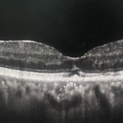
Horizontal OCT scan of a 63-year-old woman with diabetic macular edema in the right eye. When reviewing the scan, one of the intraretinal cyst (IRC) appears heart shaped. A fun scan to see just a few day's before Valentine's day.
Photographer: Kimberly Wakester, COA
Imaging device: Heidelberg
Condition/keywords: diabetic macular edema, intraretinal cyst
-
 Choroidal Melanoma
Feb 6 2025 by Virginia Gebhart
81 year old female with large pigmented collar button ciliochoroidal mass extending into the mid-vitreous cavity. Clinical exam and ultrasound finding consistent with melanoma. Due to size of tumor, pt scheduled for enucleation. CT scan of abdomen showed no evidence of metastatic disease.
Photographer: Virginia Gebhart, Retina Consultants of Carolina
Imaging device: Optos California
Condition/keywords: ciliochoroidal melanoma, collar button, melanoma
-
 New Subretinal Hemorrhage in AMD
Jan 8 2025 by Drew Mitchell
HD 1 line 100x OCT scan of a New Subretinal Hemorrhage in a established patient with AMD.
Photographer: Drew Mitchell, OCT-C
Imaging device: Zeiss Cirrus 6000
Condition/keywords: age-related macular degeneration (AMD), OCT, subretinal hemorrhage
-
 Retinal Detachment with Multiple OCT Overlays
Jan 7 2025 by Drew Mitchell
Optos 360* Color photo montage with multiple Zeiss Cirrus OCT scan overlays. Retinal Detachment with multiple breaks and a Epiretinal Membrane.
Photographer: Drew Mitchel, OCT-C
Imaging device: Optos California
Condition/keywords: ERM, macular pucker, montage, Optos, OPTOS CALIFORNIA, RD, Retinal Detachment
-
 New Choroidal Melanoma
Jan 3 2025 by Virginia Gebhart
22 year old male referred for 2nd opinion on large choroidal mass with subretinal fluid. Clinical exam and ultrasound consistent with choroidal melanoma. CT scan of orbits showed possible inflammation involving orbital fat. Pt has been on oral prednisone for 1 week, inflammation has not responded. Referred to Emory for 2nd opinion on treatment
Photographer: Virginia Gebhart
Imaging device: Optos California
Condition/keywords: melanoma
-
 New Choroidal Melanoma
Nov 7 2024 by Virginia Gebhart
83 year old female with new choroidal melanoma. Diffuse, flat tumor with orange pigment, SRF and located adjacent to ON. Pt sleeps on left side causing fluid to pool in/around the macula. Pt scheduled for brachytherapy pending CT scan results.
Photographer: Virginia Gebhart, Retina Consultants of Carolina
Imaging device: Optos California
-
 Ciliary Body Melanoma
Nov 2 2024 by Virginia Gebhart
53 year old male with a large mass behind the lens as well as prominent scleral vessels. Clinical exam and ultrasound findings consistent with melanoma. Pt will be scheduled for enucleation pending CT scan results. Edit: Sadly patient has canceled all appointments and has requested no further contact
Photographer: Virginia Gebhart, Retina Consultants of Carolina
Imaging device: Optos California
Condition/keywords: ciliary body mass, ciliary body melanoma, ciliary body tumor
-
 New Iris Melanoma
Oct 10 2024 by Virginia Gebhart
56 year old male with new amelanotic melanoma emanating from the ciliary body through the posterior iris epithelium. CT scan showed no evidence of metastatic disease. Pt scheduled for radioactive plaque and tumor biopsy
Photographer: Virginia Gebhart, Retina Consultants of Carolina
Imaging device: Samsung Galaxy
Condition/keywords: amelanotic melanoma, iris melanoma
-
 Solar Retinopathy
Mar 17 2024 by Hector Gabriel Moreno Solano, MD, MHA
OCT scan of a 65 year old male with a history of direct exposure to solar eclipse rays, visual acuity of affected eye 20/80, contralateral eye 20/25.
Photographer: Héctor Gabriel Moreno-Solano, MD, MHA
Imaging device: Revo Optopol
Condition/keywords: light toxicity, macula, solar retinopathy
-
 New Choroidal Melanoma vs Metastasis
Dec 6 2023 by Virginia Gebhart
72 year old male with possible new choroidal melanoma vs metastatic melanoma. Dome-shaped amelanotic lesion involving the fovea. Lesion was discovered during a problem visit due to sudden decreased VA (20/150). Most recent CT scan shows concern for primary lung cancer
Photographer: Virginia Gebhart
Imaging device: Topcon
Condition/keywords: amelanotic melanoma, melanoma, metastatic lesion
-
 Neovascular AMD with Ring Shaped lesions
Jul 12 2023 by Gregg T. Kokame, MD, MMM, FASRS
Horizontal OCT Scan - Neovascular AMD with Active CNV Ring shaped lesions underneath the RPE inverted U-shaped elevation
Photographer: Jaclyn Pisano
Imaging device: Zeiss Cirrus 6000
Condition/keywords: inverted u-shaped elevation, lesion, macular edema, OCT, Sub-retinal fluid, wet age-related macular degeneration (wet AMD)
-
 Neovascular AMD with Ring Shaped lesions
Jul 12 2023 by Gregg T. Kokame, MD, MMM, FASRS
Vertical OCT Scan - Neovascular AMD with Active CNV Ring shaped lesions underneath the RPE inverted U-shaped elevation
Photographer: Jaclyn Pisano
Imaging device: Zeiss Cirrus 6000
Condition/keywords: edema, lesion, OCT, Sub-retinal fluid, wet age-related macular degeneration (wet AMD)
-
 X LINKED RETINOSCHISIS
Feb 17 2023 by Ruchir Mehta, DO, DNB, FRCS
Fundus photograph and OCT scan of 8 years old male child with chronic progressive loss of vision in both eyes. His BCVA was 20/60 in both eyes. Fundus photograph showed characteristic spoke wheel pattern of foveal schisis seen in X linked Juvenile Retinoschisis. OCT showed multiple cystic spaces in foveal and perifoveal area.
Photographer: Ruchir Mehta, Mehta Superspeciality Eye Hospital, Jamnagar, Gujarat, India
Imaging device: Fundus camera
Condition/keywords: juvenile retinoschisis, OCT
-
 White Without Pressure and Peripheral Retinoschisis
Dec 29 2022 by Gulnara Islamova
Fundus Photograph and OCT scan of an 18 year-old male with peripheral retinoschisis combined with WWOP lessions .Vitreoretinal traction is not visualized
Photographer: Gulnara Islamova, CENTER ZRENIYA Medical Clinic, LLC, Chelyabinsk, Russian Federation
Imaging device: Optovue XR Avanti
Condition/keywords: peripheral retinal degeneration
-
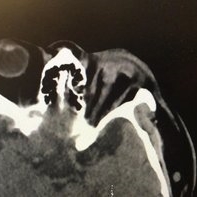 Ruptured globe on CT
Nov 19 2022 by Gareth Lema, MD, PhD
Runner meets park bench. The worst open globe I have ever seen on a CT scan. Multiple attempts at repair were made but the final visual acuity was light perception.
Photographer: Gareth Lema, MD, PhD, New York Eye and Ear of Mount Sinai
Imaging device: CT Scan
Condition/keywords: CT scan, open globe injury
-
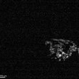
 Intraocular Foreign Body
Nov 8 2022 by pedro fernandes souza neto
Intraocular foreign body after stone trauma. CT scan image showing intraocular foreign body location.
Photographer: Pedro Fernandes, Federal University of Bahia
Condition/keywords: intraocular foreign body, trauma
-
 Floating "angle" in Vitreous
Sep 7 2022 by Stephanie Moolman
OCT scan of a vitreous floater in a 42- year-old male after anti-VEGF injection.
Photographer: Stephanie Moolman, Ophthalmic Photographer at Dr Marissa Willemse
Imaging device: Heidelberg Spectralis
Condition/keywords: black floaters, vitreous floaters
-
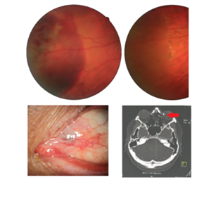 Intraocular Foreign Body
Jul 18 2022 by Nelson Chamma Capelanes, MD
Intraocular foreign body after stone trauma. Foreign body is found in the choroid. - Fundus image on the upper left: one day after the trauma showing subretinal and intraretinal hemorrhage - Fundus image on the upper right: 40 days after laser photocoagulation. - Lower left image: 30 days after the trauma, showing part of the foreign body in the nasal region. - Lower right image showing CT scan and intraocular foreign body location.
Photographer: Nelson Chamma Capelanes, Promacula Indaiatuba, Brazil
Imaging device: Canon CX-2
Condition/keywords: intraocular foreign body
 Loading…