-
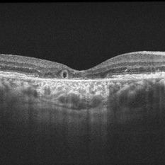
 EDI OCT Detachment With No Subretinal Fluid
Jun 29 2013 by Jason S. Calhoun
A 38-year-old male came in with blurred vision in the left eye. VA is 20/30. Notice a defect inferior of his central vision. Did an fluorescien angiogram to determine an RPE with no subretinal fluid. Also OCT confirms. Patient was injected with Avastin.
Photographer: Jason S. Calhoun, Mayo Clinic Jacksonville, Florida
Imaging device: TOPCON TRC 50-EX/CIRRUS HD OCT
Condition/keywords: central serous retinopathy (CSR), retinal pigment epithelium (RPE) detachment
-
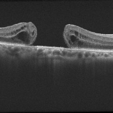 EDI OCT Macular Hole
Jun 29 2013 by Jason S. Calhoun
Enhanced depth imaging OCT shows a macular hole with no traction on middle aged female.
Photographer: Jason S. Calhoun, Mayo Clinic Jacksonville, Florida
Imaging device: TOPCON TRC 50-EX/CIRRUS HD OCT
Condition/keywords: macular hole
-
---thumb.jpeg/image-square;max$300,300.ImageHandler) Macular Traction 1
Macular Traction 1
Jul 14 2013 by Jason S. Calhoun
Macular traction with decreased vision. Patient was injected with Jetrea and will return 2-weeks for follow up.
Photographer: Jason S. Calhoun, Department of Ophthalmology, Mayo Clinic Jacksonville, Florida
Imaging device: ZEISS OCT CIRRUS
Condition/keywords: macular traction
-
---thumb.jpeg/image-square;max$300,300.ImageHandler) Macular Traction 2
Macular Traction 2
Jul 14 2013 by Jason S. Calhoun
Macular traction with decreased vision. Patient was injected with Jetrea and will return 2-weeks for follow up.
Photographer: Jason S. Calhoun, Department of Ophthalmology, Mayo Clinic Jacksonville, Florida
Imaging device: ZEISS OCT CIRRUS
Condition/keywords: macular traction
-
 EDI-Optic Neuritis/Neuroretinitis
Jul 15 2013 by Jason S. Calhoun
Patient with some loss of vision in his left eye last week, was seen by eye MD and referred for eval. Patient also complained of pain in left eye with eye movement. VA was 20/400 in the left eye. Fundus photo and HD-OCT imaging show optic nerve swelling and fluid underneath the retina. A neuro-ophthalmologist will be consulted for further evaluation.
Photographer: Jason S. Calhoun, Department of Ophthalmology, Mayo Clinic Jacksonville, Florida
Imaging device: ZEISS OCT CIRRUS
Condition/keywords: neuroretinitis, optic neuritis
-
 EDI-Optic Neuritis/Neuroretinitis
Jul 15 2013 by Jason S. Calhoun
Patient with some loss of vision in his left eye and was seen for an evaluation. Patient also complained of pain in left eye with eye movement. VA was 20/400 in the left eye. Fundus photo and HD-OCT imaging show optic nerve swelling and fluid underneath the retina. A neuro-ophthalmologist will be consulted for further evaluation.
Photographer: Jason S. Calhoun, Department of Ophthalmology, Mayo Clinic Jacksonville, Florida
Imaging device: ZEISS OCT CIRRUS
Condition/keywords: neuroretinitis, optic neuritis
-
 Outer-Retinal-Tubulation
Jun 27 2013 by Jason S. Calhoun
Patient with a history of wet macular degeneration and glaucoma in both eyes. VA is 20/50, right eye, 20/80, left eye. Patient is treated with Eylea in both eyes. Enhanced depth imaging OCT reveals a small like form of a cyst which in fact isn't a cyst at all. This is called outer retinal tubulation in which degenerating photo-receptors may become arranged in a circular or ovoid fashion. This is sometimes misdiagnosed as cystic changes in the retinal pigment epithelium or sub-retinal fluid.
Photographer: Jason S. Calhoun, Mayo Clinic Jacksonville, Florida
Imaging device: ZEISS OCT CIRRUS
Condition/keywords: optical coherence tomography (OCT)
A project from the American Society of Retina Specialists