-
 Chronic Serous Central Choroidopathy
Oct 15 2025 by Vicente Nicanor Mancilla Guerrero
Color mosaic fundus image of a 60-year-old male patient with CSC recurrences, with the presence of retinal tapestry atrophy with gravitational pattern.
Photographer: Vicente Mancilla G, Ophthalmic Medical Technologist
Imaging device: Canon cx-1
Condition/keywords: chronic central serous chorioretinopathy (CSCR)
-
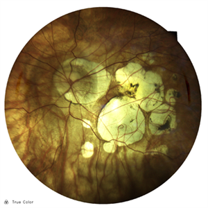 Myopic Degeneration
Oct 23 2025 by Vicente Nicanor Mancilla Guerrero
Confocal laser retinography of a female patient with -20.00Dp myopia. Typical findings of myopic retinopathy can be seen.
Photographer: Vicente mancilla G, Ophthalmic Medical Technologist
Imaging device: Compass Centervue
Condition/keywords: high myopia
-
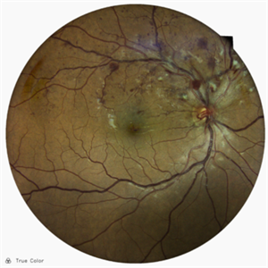 Superior Temporal Venous Branch Occlusion
Oct 23 2025 by Vicente Nicanor Mancilla Guerrero
Confocal laser retinography of the right eye of a 45-year-old female patient with hypertension (+). Venous dilation is evidenced together with the presence of flame hemorrhages and cottony spots in the upper temporal arch.
Photographer: Vicente Mancilla G, Medical Technologist in Ophthalmology
Imaging device: Compass CenterVue
Condition/keywords: branch retinal vein occlusion (BRVO)
-
 Lower Nasal Venous Branch Obstruction
Nov 20 2025 by Vicente Nicanor Mancilla Guerrero
Early fluorescein angiography of the left eye of a 57-year-old patient who presents with a long-standing inferior nasal venous branch occlusion. Dilation and tortuosity can be seen in the lower arch, along with the classic areas of retinal ischemia.
Photographer: Vicente Mancilla Guerrero, Ophthalmic Medical Technologist
Imaging device: Canon cx-1
Condition/keywords: branch retinal vein occlusion (BRVO)
-
 Atypical Grade Foveal Hypoplasia
Aug 9 2026 by Vicente Nicanor Mancilla Guerrero
Left eye line report of a female patient with suspected ocular albinism, showing an atypical grade of bilateral foveal hypoplasia according to the Thomas classification. The most prominent findings are the persistence of inner retinal layers, reduction of the foveal contour, and clear disruption of the ellipsoid zone at the foveal level. As an additional finding, fundus photography revealed a hypopigmented retinal background.
Photographer: Vicente Mancilla G, Medical Technologist in Ophthalmology
Imaging device: Angiovue, Optovue OCT
Condition/keywords: albinism, foveal hypoplasia, hypo
-
 Paraneoplastic Optic Neuritis
Aug 10 2026 by Vicente Nicanor Mancilla Guerrero
Male patient diagnosed with paraneoplastic optic neuritis of the left eye. This is a rare phenomenon, usually associated with small-cell carcinomas. It is believed that tumor-associated antigens such as CRMP-5 IgL generate antibodies that cross-react with neuronal and glial proteins; aside from neuritis, they can trigger the neuromyelitis optica spectrum. In this patient, this neuritis progressed to complete optic nerve atrophy, as this is a blind eye.
Photographer: Vicente Mancilla G, Medical technologist specialized in ophthalmology
Imaging device: Compass Centervue iCare
Condition/keywords: optic neuritis, paraneoplastic
-
 Proliferative Diabetic Retinopathy
Aug 31 2026 by Vicente Nicanor Mancilla Guerrero
Fundus examination of a young female patient with type 1 diabetes mellitus, with a diabetes duration of over 10 years, currently diagnosed with proliferative diabetic retinopathy. Laser spots can be seen in the periphery, multiple hemorrhagic foci in all four quadrants, and exudation.
Photographer: Vicente Mancilla G, Ophthalmic Medical Technologist
Imaging device: Compass CenterVue
Condition/keywords: diabetes
-
 Chimney Smoke
Oct 21 2026 by Vicente Nicanor Mancilla Guerrero
A 30-year-old male patient presents with sudden vision loss in the left eye. A diagnosis of central serous chorioretinopathy (CSCR) is suspected. In the late-phase fluorescein angiography image, we can observe the classic “smokestack” sign, which appears as the dye enters the serous detachment. Treatment will depend on the course of the condition over a period of 1 to 3 months.
Photographer: Vicente Mancilla G, Medical technologist specialized in ophthalmology
Imaging device: TOPCON TRC-50DX
Condition/keywords: central serous chorioretinopathy (CSCR)

A project from the American Society of Retina Specialists