-
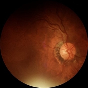
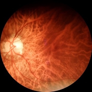
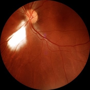
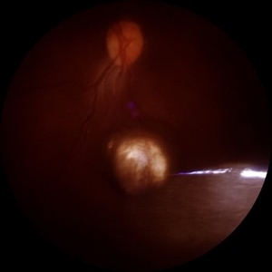
 Retinocoroiditis Inactiva Por Toxoplasmosis
Apr 28 2025 by Paulina Araujo
Fundus photography demonstrates a 2-disc-diameter chorioretinal scar in the superior temporal arcade, consistent with inactive toxoplasmic retinochoroiditis. The lesion exhibits pigmented borders and central atrophy, with adjacent splinter hemorrhages and vascular sheathing. No vitreous inflammation or active satellite lesions are present.
Photographer: Paulina D.Araujo Martínez, Asociación para Evitar la Ceguera en México I.A.P., Hospital Dr Luis Sánchez Bulnes.
Condition/keywords: toxoplasmosis chorioretinitis
-
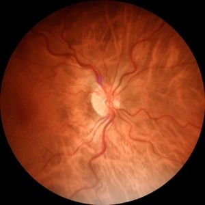
 Atrophy
Jun 4 2025 by Paulina Araujo
The fundus photograph captures the central 55 degrees of the right eye, revealing alpha and beta peripapillary atrophy.
Photographer: Paulina D.Araujo Martínez, Asociación para Evitar la Ceguera en México I.A.P., Hospital Dr Luis Sánchez Bulnes.
Condition/keywords: atrophy
-
 Tractional Retinal Detachment
Jun 4 2025 by Paulina Araujo
The 55-degree central fundus photograph of the right eye reveals a thickened and opacified hyaloid exerting traction on the optic disc and posterior pole of the retina, along with hard exudates and microaneurysms consistent with advanced proliferative diabetic retinopathy.
Photographer: Paulina D.Araujo Martínez, Asociación para Evitar la Ceguera en México I.A.P., Hospital Dr Luis Sánchez Bulnes.
Condition/keywords: tractional retinal detachment
-
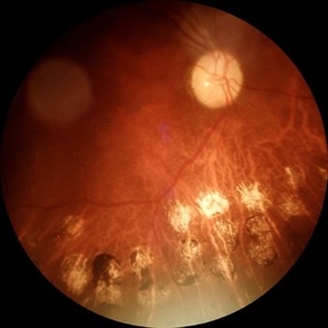
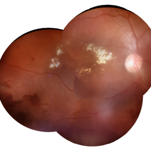 Macular Edema
Jun 4 2025 by Paulina Araujo
The composite fundus photograph of the right eye demonstrates circinate hard exudates in the thickened macular area, along with flame-shaped intraretinal hemorrhages along the inferior temporal arcade.
Photographer: Paulina D.Araujo Martínez, Asociación para Evitar la Ceguera en México I.A.P., Hospital Dr Luis Sánchez Bulnes.
Condition/keywords: macular edema
-
 Tessellated Fundus
Jun 4 2025 by Paulina Araujo
The 55-degree central fundus photograph of the left eye reveals a tessellated fundus appearance consistent with high myopia.
Photographer: Paulina D.Araujo Martínez, Asociación para Evitar la Ceguera en México I.A.P., Hospital Dr Luis Sánchez Bulnes.
Condition/keywords: Tessellated fundus
-
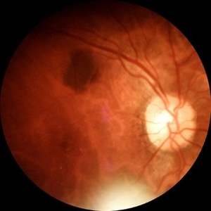
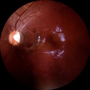 Choroidal Rupture
Jun 4 2025 by Paulina Araujo
The 55-degree central fundus photograph of the left eye reveals a choroidal rupture in the nasal parafoveal area secondary to blunt ocular trauma.
Photographer: Paulina D.Araujo Martínez, Asociación para Evitar la Ceguera en México I.A.P., Hospital Dr Luis Sánchez Bulnes.
Condition/keywords: choroidal rupture
-
 Laser Marks
Jun 4 2025 by Paulina Araujo
The 55-degree central fundus photograph of the right eye shows evidence of old laser marks along the inferior temporal arcade.
Photographer: Paulina D.Araujo Martínez, Asociación para Evitar la Ceguera en México I.A.P., Hospital Dr Luis Sánchez Bulnes.
Condition/keywords: Attached retina with Endolaser marks, laser
-
 Bear Tracks (CHRPE)
Jun 4 2025 by Paulina Araujo
The 55-degree fundus photograph of the left eye shows bear tracks along the inferior temporal arcade.
Photographer: Paulina D.Araujo Martínez, Asociación para Evitar la Ceguera en México I.A.P., Hospital Dr Luis Sánchez Bulnes.
Condition/keywords: bear tracks, congenital hypertrophy of the retinal pigment epithelium (CHRPE)
-
 Myelinated Nerve Fibers
Jun 4 2025 by Paulina Araujo
The 55-degree central fundus photograph of the left eye reveals myelination of the nerve fiber layer along the inferior nasal arcade.
Photographer: Paulina D.Araujo Martínez, Asociación para Evitar la Ceguera en México I.A.P., Hospital Dr Luis Sánchez Bulnes.
Condition/keywords: myelinated nerve fibers
-
 Choroidal Nevus
Jun 4 2025 by Paulina Araujo
The 55-degree central fundus photograph of the right eye reveals a choroidal nevus measuring 0.5 disc diameters along the superior temporal arcade.
Photographer: Paulina D.Araujo Martínez, Asociación para Evitar la Ceguera en México I.A.P., Hospital Dr Luis Sánchez Bulnes.
Condition/keywords: Choroidal nevus
-
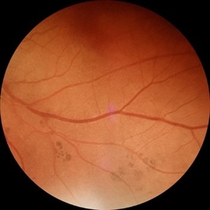
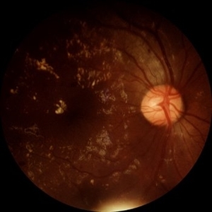 Diabetic Retinopathy
Jun 4 2025 by Paulina Araujo
The 55-degree central fundus photograph of the right eye demonstrates numerous hard exudates, dot intraretinal hemorrhages, and microaneurysms.
Photographer: Paulina D.Araujo Martínez, Asociación para Evitar la Ceguera en México I.A.P., Hospital Dr Luis Sánchez Bulnes.
Condition/keywords: diabetic retinopathy
-
 Hypertensive Retinopathy
Jun 4 2025 by Paulina Araujo
The 55-degree central fundus photograph of the right eye reveals vascular tortuosity, generalized arteriolar narrowing with a vein-to-artery ratio of 3:1, along with Guist and Bonnet signs.
Photographer: Paulina D.Araujo Martínez, Asociación para Evitar la Ceguera en México I.A.P., Hospital Dr Luis Sánchez Bulnes.
Condition/keywords: hypertensive retinopathy
-
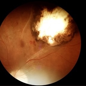
 Tuberculoma
Jun 4 2025 by Paulina Araujo
The 55-degree central fundus photograph of the left eye reveals an elevated, nodular whitish choroidal lesion along the inferior temporal arcade, consistent with a tuberculoma.
Photographer: Paulina D.Araujo Martínez, Asociación para Evitar la Ceguera en México I.A.P., Hospital Dr Luis Sánchez Bulnes.
Condition/keywords: Choroidal-tuberculoma
-
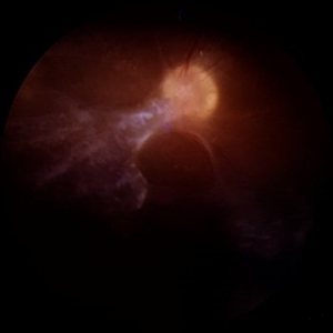
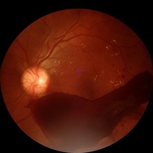 Subhyaloid Hemorrhage
Jul 16 2025 by Paulina Araujo
55-degree central fundus photograph of the left eye (OS) shows a prominent subhyaloid hemorrhage in the inferior posterior pole, displaying a characteristic 'boat-shaped' appearance with well-defined margins and dark red coloration.
Photographer: Paulina D.Araujo Martínez, Asociación para Evitar la Ceguera en México I.A.P., Hospital Dr Luis Sánchez Bulnes.
Condition/keywords: subhyaloid hemorrhage
A project from the American Society of Retina Specialists