-
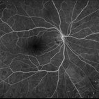
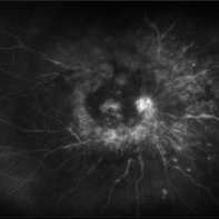 Posterior Uveitis with Macular Edema
Jul 9 2024 by Korey Starkey
Ultra-wide field angiography of a 70 year old female with cystoid macular edema secondary to posterior uveitis. Patient's vision was Dcc20/200 at time of visit.
Photographer: Korey Starkey
Imaging device: Optos
Condition/keywords: cystoid macular edema (CME), fluorescein angiogram (FA), FLUORESCEIN ANGIOGRAPHY, hyperfluorescence, posterior uveitis, ULTRA WIDE FIELD, ultra-widefield image, vitreous debris
-
 Panuveitis
Jul 12 2024 by Korey Starkey
Ultra widefield Optos FA of 59 year old female presents with panuveitis in both eyes. Patients vision was VA OS: Dcc20/60-2 at time of visit.
Photographer: Korey Starkey
Imaging device: Optos
Condition/keywords: FLUORESCEIN ANGIOGRAPHY, hyperfluorescence, Optos, Panuveitis, ultra-wide field imaging, Uveitis
-
 Active Proliferative Diabetic Retinopathy
Jul 12 2024 by Korey Starkey
Fluorescein angiogram performed on 35 year old female with active proliferative diabetic retinopathy. Patient has peripapillary vascular loop and history of PRP treatment in both eyes. Patients left eye vision measured at Dcc20/200-1 at this visit.
Photographer: Korey Starkey
Imaging device: Optos
Condition/keywords: FLUORESCEIN ANGIOGRAPHY, hyperfluorescence, laser scarring, Optos, proliferative diabetic retinopathy (PDR), sea fan, ultra-wide field imaging, vascular loop
-
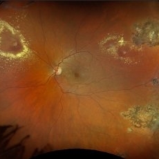
 Choroidal Hemangioma
Jul 30 2024 by Korey Starkey
ICG image of a77 year-old female with choroidal hemangioma. The physician states the hypercyanesence in the right eye is consistent with hemangioma but no typical late washout observed. He also notes high internal reflectivity make hemangioma possible. Patients vision at time of imaging VA OD: sc20/200 PH20/60-1; plan to follow patient at 6 month intervals at this time.
Photographer: Korey Starkey
Imaging device: Optos
Condition/keywords: Choroidal Hemangioma, Fluorescein angiography, indocyanine green (ICG) angiography, Optos
-
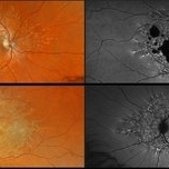 Stargardt Disease
Aug 27 2024 by Korey Starkey
Ultra wide-field fundus photograph and fundus autofluorescence of a 49-year-old male. Initial visit imaging.
Photographer: Korey Starkey
Imaging device: Optos
Condition/keywords: fundus autofluorescence (FAF), fundus photograph, Optos, Stargardt disease, ultra-wide field imaging
-
 Central Retinal Vein Occlusion
Sep 27 2024 by Korey Starkey
Fluorescein angiogram of a 75 year old patient with central retinal vein occlusion. FA shows areas of patchy ischemia and petaloid leakage. Patient is being treated with anti-vegf treatments at this time.
Photographer: Korey Starkey
Condition/keywords: central retinal vein occlusion (CRVO), FLUORESCEIN ANGIOGRAPHY, ischemia, macular edema, petaloid leakage, ultra-widefield image
-
 NVI
NVI
Oct 24 2024 by Korey Starkey
Iris FA of a 74 year old male with neovascularization of the iris. Noted mild activity of NVI at the superior pupillary margin, recommending observation at time of visit.
Photographer: Korey Starkey
Imaging device: Heidelberg Spectralis
Condition/keywords: FA, Heidelburg Spectralis, Iris, iris fluorescein angiogram, neovascularization of iris (NVI), smokestack
-
 PFO Bubbles
Oct 24 2024 by Korey Starkey
Slit lamp photograph of a 23 year old female with PFO bubbles inferiorly in the AC. Discussed surgical intervention to remove PFO from AC and vitreous cavity in future.
Photographer: Korey Starkey
Imaging device: Slit lamp camera
Condition/keywords: anterior chamber, PFO, slit lamp photography, submacular perfluorocarbon liquid (PFO)
-
 Diabetic Retinopathy
Nov 20 2024 by Korey Starkey
64 year old female being monitored for moderate-severe diabetic retinopathy.
Photographer: Korey Starkey
Condition/keywords: capillary nonperfusion, FA, FLUORESCEIN ANGIOGRAPHY, microaneurysms, nonproliferative diabetic retinopathy, Optos, OPTOS CALIFORNIA, tortuous vessels
-
 Chronic CRVO
Dec 12 2024 by Korey Starkey
Fluorescein Angiography of a 62 year-old man with chronic central retinal vein occlusion. Vision is 20/200.
Photographer: Korey Starkey
Imaging device: Optos
Condition/keywords: capillary nonperfusion, central retinal vein occlusion (CRVO), FLUORESCEIN ANGIOGRAPHY, ischemia, microaneurysms, Optos
-
 Vitreous Prolapse
Jan 28 2025 by Korey Starkey
Slit lamp image of a 62-year-old patient presented at first visit with vitreous prolapse due to mechanical complications from IOL placement. IOP was being managed with drops, vision was 20/20, patient opted for surgery due to constant haze in vision.
Photographer: Korey Starkey
Imaging device: Slit lamp camera
Condition/keywords: slit lamp photo, vitreous prolapse
-
 Epicapsular Stars
Jan 28 2025 by Korey Starkey
Epicapsular stars and cataract noted in natural lens of 68-year-old patient.
Photographer: Korey Starkey
Imaging device: Slit lamp camera
Condition/keywords: cataract, chicken tracks, epicapsular stars, slit lamp photography
-
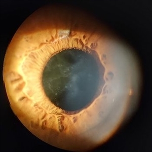
 Iris Melanoma
Jan 28 2025 by Korey Starkey
Slit-lamp image of 90-year-old patient with iris melanoma and new hemorrhage affecting the right eye. Patient re-presented after nearly 1 year, now seeking treatment. Given iris location of tumor, multiple clock hours of iris involved, and increase in size of the known malignant transformation; safest approach was enucleation.
Photographer: Korey Starkey
Imaging device: Slit lamp camera
Condition/keywords: anterior chamber, hemorrhage, iris melanoma, slit lamp photo
-
 Iris Nevus
Jan 28 2025 by Korey Starkey
Slit-lamp image of an 89-year-old patient with an iris nevus. Nevus appeared stable on exam, will continue to monitor.
Photographer: Korey Starkey
Imaging device: Slit lamp camera
Condition/keywords: ectropion uveae, iris nevus, slit lamp photo
-
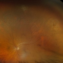
 Retinal Detachment and Lattice Degeneration
Mar 25 2025 by Korey Starkey
26 year-old patient presented at first visit with rhegmatogenous macula involving retinal detachment of the left eye. Underwent prompt surgical repair. Both eyes also present with lattice degeneration with atrophic holes.
Photographer: Korey Starkey
Condition/keywords: atrophic retinal hole, fundus photography, lattice degeneration, montage photo, Optos, OPTOS CALIFORNIA RGB, retinal detachment, retinal holes, rhegmatogenous retinal detachment, ultra-wide field imaging
-
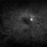
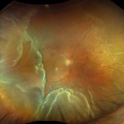
 Retinal Vasculitis
Mar 26 2025 by Korey Starkey
41 year-old patient presents with vascular FA findings of occlusive vasculitis with four quadrant Kyrieleis plaques OU showcases a possibly rare but reported atypical presentation of Behcet's Syndrome.
Photographer: Korey Starkey
Imaging device: Optos
Condition/keywords: Behcet's Disease, FA early phase, Fundus Fluorescein Angiography, Optos, retinal vasculitis, ultra-wide field imaging, venous beading
-
 Retinal Vasculitis
Mar 26 2025 by Korey Starkey
41 year-old patient presents with vascular FA findings of occlusive vasculitis with four quadrant Kyrieleis plaques OU showcases a possibly rare but reported atypical presentation of Behcet's Syndrome.
Photographer: Korey Starkey
Imaging device: Optos
Condition/keywords: FA early phase, Fundus Fluorescein Angiography, ischemia, Optos, retinal vasculitis, ultra-wide field imaging, venous beading
-
 ROP
ROP
Mar 26 2025 by Korey Starkey
9 month old patient presents today with Retinopathy of Prematurity in both eyes. Patient was born at gestational age of 25 weeks 2 days, 940g. Left eye presents with vitreoretinal traction and peripheral VH with regressed stage 3 and persistent stage 2 disease.
Photographer: Korey Starkey
Imaging device: Optos
Condition/keywords: retinopathy of prematurity stage 2, rop, stage 3, vitreoretinal traction, vitreous hemorrhage
-
 Ozurdex in AC
Apr 1 2025 by Korey Starkey
90-year-old patient with an Ozurdex implant that migrated into the AC and with the cornea decompensating. Patient recommended for urgent surgery to remove implant. Vision OD at this visit was CF @ 2ft, most recent visit vision is 20/400, PH 20/25.
Photographer: Korey Starkey
Imaging device: Topcon
Condition/keywords: anterior chamber, corneal decompensation, external, external photography, Ozurdex implant, Topcon
-
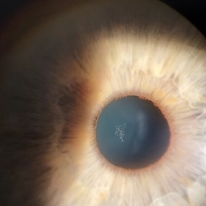
 Iris Discoloration
Apr 1 2025 by Korey Starkey
4 year-old patient sent for genetic testing to rule out possibility of Waardenburg syndrome with Hirschsprung disease. Left eye iris has no discoloration present, vision in both eyes is 20/40.
Photographer: Korey Starkey
Imaging device: Topcon
Condition/keywords: external, external photography, iris, topcon
-
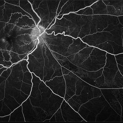
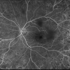
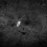
 Shunt Vessels
Apr 1 2025 by Korey Starkey
62-year-old patient presented with stable CRVO in the left eye. FA performed that day shows delayed AV transit is present, this has compensated with shunt vessels at the disc. However there is no evidence of active leakage. OS vision 20/25.
Photographer: Korey Starkey
Imaging device: Topcon
Condition/keywords: central retinal vein occlusion (CRVO), fundus photograph, optic nerve, shunts vessels, Topcon
-
 Central Retinal Vein Occlusion With Waldenstroms macroglobulinemia
Jun 18 2025 by Korey Starkey
64-year-old patient presents with CRVO with secondary macular edema in both eyes. Venous beading present in 2/4 quadrants OU. Patient diagnosed with Waldenstroms macroglobulinemia, found on SPEP and bone marrow biopsy. Treatment recommended of anti-vegF intravitreal injections OU.
Photographer: Korey Starkey
Imaging device: Optos
Condition/keywords: attenuated vessels, central retinal vein occlusion (CRVO), CRVO, FA early phase, FLUORESCEIN ANGIOGRAPHY, macular edema, Optomap, OPTOS CALIFORNIA, severe NPDR, venous beading, Waldenstroms macroglobulinemia
-
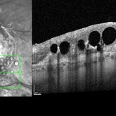 Subretinal PFO
Jun 18 2025 by Korey Starkey
86-year-old patient had history for retinal detachment surgery x2 and intraocular injections for AMD performed elsewhere. Left eye has PVR developing and subretinal PFO. Due to guarded vision, opting to defer any further treatment at this time.
Photographer: Korey Starkey
Imaging device: Heidelberg
Condition/keywords: AMD, Heidelburg Spectralis, OCT, PFO, PVR, retinal detachment, silicone oil
-
 Ocular Ischemic Syndrome
Jun 18 2025 by Korey Starkey
58-year-old patient with OIS in both eyes. Patient has had PRP in the past, however, presence of NVD with peripheral nonperfusion remains despite PRP.
Photographer: Korey Starkey
Imaging device: Optos
Condition/keywords: DME, FA early phase, fluorescein angiogram (FA), NVD, ocular ischemic syndrome, ois, Optos, peripheral retinal nonperfusion
-
 Ocular Ischemic Syndrome
Jun 18 2025 by Korey Starkey
58-year-old patient with OIS and Hollenhorst plaque.
Photographer: Korey Starkey
Imaging device: Optos
Condition/keywords: capillary nonperfusion, fluorescein angiogram (FA), hollenhorst plaque, NVD, ocular ischemic syndrome, Optos
-
 Retinal Aneurysms
Aug 6 2025 by Korey Starkey
54 year-old patient presents with scattered peripheral aneurysms with exudates. FA was performed showing peripheral nonperfusion and aneurysms. Treated patient with PRP and focal laser to aneurysms and continued observation.
Photographer: Kore Starkey
Imaging device: Optos
Condition/keywords: aneurysm, branch retinal vein occlusion (BRVO), chorioretinal scar, circinate ring, exudates, fundus photography, lesion, Optos, retinal aneurysms
-
 Horseshoe Retinal Tear
Aug 6 2025 by Korey Starkey
80 year-old patient presented with HSRT without detachment in the left eye and macula-off detachment in the right eye. Scheduled patient for prompt surgical repair OD and same day laser retinopexy OS to reduce risk of retinal detachment.
Photographer: Korey Starkey
Imaging device: Optos
Condition/keywords: color fundus photograph, fundus photography, horseshoe tear, Optos
-
 Total Retinal Detachment
Aug 6 2025 by Korey Starkey
59 year-old patient presents with total retinal detachment at first visit in OD. Recommending prompt surgical intervention.
Photographer: Korey Starkey
Imaging device: Optos
Condition/keywords: color fundus photograph, Optos, retinal detachment, total retinal detachment
-
 Retinal Arteriovenous Malformation
Oct 7 2025 by Korey Starkey
55 year-old patient presents with retinal arteriovenous malformation in the left eye and BRVO w/retinal neovascularization. Patient is asymptomatic. No edema or treatment necessary today, signs of old RVO with MAs along inferior arcade and dot heme.
Photographer: Korey Starkey
Imaging device: Topcon
Condition/keywords: branch retinal vein occlusion (BRVO), fundus photography, inferior arcade, microaneurysms, retinal arteriovenous malformations, retinal neovascularization, Topcon
-
 Retinal Detachment with Macular Hole
Nov 10 2025 by Korey Starkey
A 58 year-old female presented with re-detached retina through macular hole. Planned for surgical intervention.
Photographer: Korey Starkey
Imaging device: Optos
Condition/keywords: gas bubble, intermediate uveitis, macular hole, pars plana vitrectomy (PPV), retinal detachment, scleral buckle, traction detachment
A project from the American Society of Retina Specialists