-
 Isolated Choroidal Melanocytosis
Oct 27 2019 by John S. King, MD
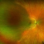
23-year-old white female consulted for a large pigmented choroidal lesion in the right eye. Healthy, no history of glaucoma, 20/20 OU without scleral pigment changes; large, flat pigmented choroidal lesion that is sectoral (temporally) and pigment appears to spare the major choroidal vessels. On OCT (not shown) there is mildly increased choroidal thickening in the area of the lesion.
Photographer: Shelly Blair
Imaging device: Optos CA
Condition/keywords: choroidal melanocytosis, ocular melanocytosis
-
 Isolated Choroidal Melanocytosis - Montage
Oct 27 2019 by John S. King, MD
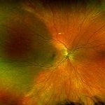
23-year-old white female consulted for a large pigmented choroidal lesion in the right eye. Healthy, no history of glaucoma, 20/20 OU without scleral pigment changes; large, flat pigmented choroidal lesion that is sectoral (temporally) and pigment appears to spare the major choroidal vessels. On OCT (not shown) there is mildly increased choroidal thickening in the area of the lesion.
Photographer: Shelly Blair
Imaging device: Optos CA
Condition/keywords: choroidal melanocytosis, ocular melanocytosis
A project from the American Society of Retina Specialists