-
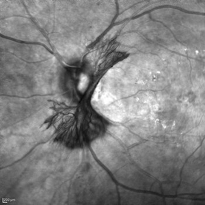 "NVD Flower"
Oct 20 2023 by Daniel Davis, OCT-C
Infrared image of NVD (52F)
Imaging device: Heidelberg Spectralis
Condition/keywords: neovascularization of the disc (NVD)
-
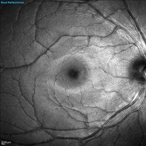 PAMM-BLUE
Nov 29 2023 by Daniel Davis, OCT-C
Blue reflectance image of a 30 yo female with PAMM OD.
Photographer: Daniel Davis, OCT-C
Imaging device: Heidelberg Spectralis
Condition/keywords: blue reflectance
-
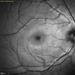 PAMM-GREEN
Nov 29 2023 by Daniel Davis, OCT-C
Green reflectance of a 30 yo female with PAMM OD.
Photographer: Daniel Davis, OCT-C
Imaging device: Heidelberg Spectralis
Condition/keywords: red-free
-
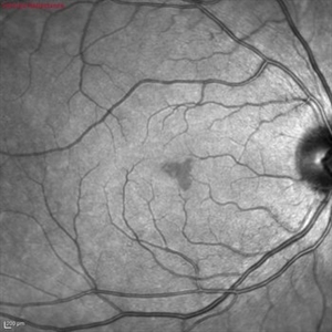 PAMM-IR
Nov 29 2023 by Daniel Davis, OCT-C
Infrared fundus of a 30 yo female with PAMM OD.
Photographer: Daniel Davis, OCT-C
Imaging device: Heidelberg Spectralis
Condition/keywords: infrared image
-
 PAMM-MultiColor
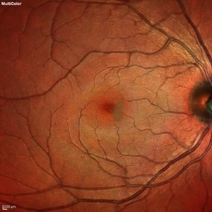
Nov 29 2023 by Daniel Davis, OCT-C
Multi-color fundus of a 30 yo female with PAMM OD.
Photographer: Daniel Davis, OCT-C
Imaging device: Heidelberg Spectralis
Condition/keywords: multicolor
-
 PAMM-OCTA
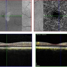
Nov 29 2023 by Daniel Davis, OCT-C
OCT-A of a 30 yo female with PAMM OD.
Photographer: Daniel Davis, OCT-C
Imaging device: Heidelberg Spectralis
Condition/keywords: OCTA, paracentral acute middle maculopathy
-
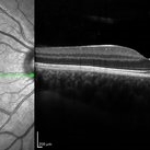
Paracentral Acute Middle Maculopathy
Nov 29 2023 by Daniel Davis, OCT-C
30 yo female OCT with Paracentral Acute Middle Maculopathy (PAMM) OD VA OD: sc20/60+1
Condition/keywords: OCT, PAMM
-
 Acute Macular Neuroretinopathy
Mar 25 2024 by Daniel Davis, OCT-C
18 yo female presenting with hazy vison for 2-3 weeks. VA OD: sc20/20 VA OS: sc20/20 Infrared imaging showed dark gray, petalloid, perifoveal lesions and OCT shows focal signal reduction of the Inner Segment / Outer Segment junction. Elects to observe.
Photographer: Daniel Davis, OCT-C, The Retina Institute, St. Louis
Imaging device: Optos California SWL
Condition/keywords: acute macular neuroretinopathy
-
 Choroidal Melanoma with Serous Retinal Detachment
Dec 20 2024 by Daniel Davis, OCT-C
67 year old male presenting with large pigmented choroidal mass with serous retinal detachment.
Photographer: Daniel Davis, OCT-C, The Retina Institute
Imaging device: Optos California
Condition/keywords: Retina detachment
-
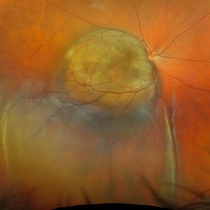
 Astrocytic Hamartoma
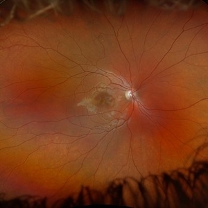
Feb 27 2025 by Daniel Davis, OCT-C
Color fundus photo of 55-year-old female with Astrocytic Hamartoma in association with tuberous sclerosis. No treatment options available, benign. Other findings include; Posterior Vitreous Detachment, Vitreous Hemorrhage, Hereditary Retinal Dystrophy, Vitreous Opacities, Hypertensive Retinopathy.
Photographer: Daniel Davis, OCT-C
Imaging device: Optos California
Condition/keywords: color fundus photograph
-
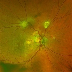
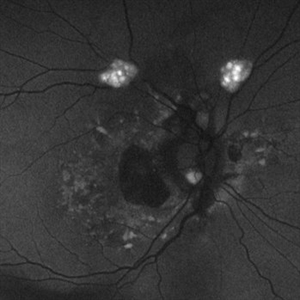 Astrocytic Hamartoma
Feb 27 2025 by Daniel Davis, OCT-C
Fundus autofluorescence photo of 55-year-old female with astrocytic hamartoma in association with tuberous sclerosis. No treatment options available, benign. Other findings include; Posterior Vitreous Detachment, Vitreous Hemorrhage, Hereditary Retinal Dystrophy, Vitreous Opacities, Hypertensive Retinopathy.
Photographer: Daniel Davis, OCT-C
Imaging device: Optos California
Condition/keywords: astrocytic hamartoma, fundus autofluorescence (FAF)
A project from the American Society of Retina Specialists