-
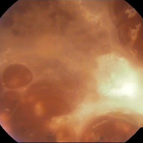 Proliferative Diabetic Retinopathy
Mar 25 2013 by Ratimir Lazic, MD, PhD
Color fundus photography of a 62- year-old diabetic patient. Severe fibrous proliferations with traction retinal detachment can be seen. Pars plana vitrectomy was preformed on that eye.
Photographer: Marko Lukic, MD
Imaging device: Zeis Visucam Lite 2
Condition/keywords: neovascularization (NV)
-
---thumb.jpg/image-square;max$300,300.ImageHandler) Proliferative Diabetic Retinopathy
Proliferative Diabetic Retinopathy
Mar 25 2013 by Ratimir Lazic, MD, PhD
Color fundus photography of a 62- year-old diabetic patient. Fibrous proliferations along upper temporal branch and posterior pole with no traction on retina can be seen. Suspected neovascularization nasal from PNO.
Photographer: Marko Lukic, MD
Imaging device: Zeis Visucam Lite 2
Condition/keywords: neovascularization (NV)
-
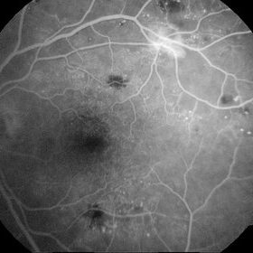 Neovascularization - RDP
Jun 29 2014 by Ratimir Lazic, MD, PhD
A FAG image of a 84-year-old female. Late venous phase of the left eye. Hyperfloercent area in upper temporal quadrant represents NVE. Many hyperflorescent dots can be seen. Few hypoflorescent areas are deep retinal hemorrhages.
Photographer: Marko Lukic, University Eye Clinic Svjetlost
Imaging device: Zeis Visucam Lite 2
Condition/keywords: neovascularization (NV), neovascularization elsewhere (NVE)
-
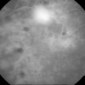 Neovascularization - RDP
Jun 29 2014 by Ratimir Lazic, MD, PhD
FAG image of a 84-year-old female. Dye leakage from the NVE can be seen.
Photographer: Marko Lukic, University Eye Clinic Svjetlost
Imaging device: Zeis Visucam Lite 2
Condition/keywords: neovascularization elsewhere (NVE)
-
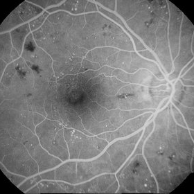 Diabetic Retinopathy
Jun 29 2014 by Ratimir Lazic, MD, PhD
A FAG image of a 84-year-old female. Diabetic changes of the posterior pole and midperipheral retina can be seen. Mild dye leakage in macula with many hyperflorescent dots (microaneurisms) and hypoflorescent areas (intraretinal hemorrhages) can be seen.
Photographer: Marko Lukic, University Eye Clinic Svjetlost
Imaging device: Zeis Visucam Lite 2
Condition/keywords: diabetic retinopathy
-
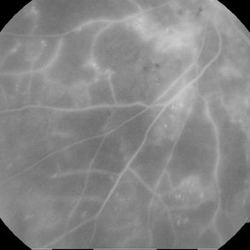 Proliferative Diabetic Retinopathy - Capillary Dropouts
Jun 29 2014 by Ratimir Lazic, MD, PhD
A FAG image of a 84-year-old female. Extensive areas of capillary dropouts of mid and peripheral retina can be noticed. Due to macular edema and capillary dropouts we reccomended combination of intravitreal antiVEGF therapy and argon laser treatment. .
Photographer: Marko Lukic, University Eye Clinic Svjetlost
Imaging device: Zeis Visucam Lite 2
Condition/keywords: anti-VEGF, capillary dropouts
A project from the American Society of Retina Specialists