-
 Toxoplasmosis
Oct 9 2019 by McGill University Health Centre
A 29-year-old immunosuppressed woman with extensive retinitis extending through the macula.
Photographer: Miguel N. Burnier, McGill University Health Center-McGill University Ocular Pathology & Translational Research Laboratory
Condition/keywords: AIDS, toxoplasmosis retinitis
-
 Toxoplasmosis
Oct 9 2019 by McGill University Health Centre
A 29-year-old immunosuppressed woman with toxoplasmosis. Gross pathology of enucleated eye with extensive retinitis.
Photographer: Miguel N. Burnier, McGill University Health Center-McGill University Ocular Pathology & Translational Research Laboratory
Condition/keywords: AIDS, gross pathology, toxoplasmosis, toxoplasmosis retinitis
-
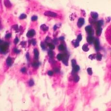 Toxoplasmosis
Oct 9 2019 by McGill University Health Centre
A 29-year-old immunosuppressed woman with toxoplasmosis. Higher power of the retina showing Toxoplasma gondii cysts.
Photographer: Miguel N. Burnier, McGill University Health Center-McGill University Ocular Pathology & Translational Research Laboratory
Condition/keywords: histopathology, retina, toxoplasma gondii cysts, toxoplasmosis
-
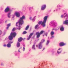 Toxoplasmosis
Oct 9 2019 by McGill University Health Centre
A 29-year-old immunosuppressed woman with toxoplasmosis. Higher power of the retina showing Toxoplasma gondii cysts.
Photographer: Miguel N. Burnier, McGill University Health Center-McGill University Ocular Pathology & Translational Research Laboratory
Condition/keywords: histopathology, retina, toxoplasma gondii cysts, toxoplasmosis
-
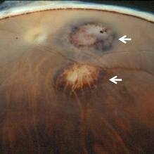 Toxoplasmic Chorioretinitis
May 18 2020 by McGill University Health Centre
Toxoplasmic chorioretinitis is caused by parasitic infection from Toxoplasma gondii. Two forms are recognized: congenital and acquired. Congenital toxoplasmic chorioretinitis occurs because the infection is transplacental: T. gondii is among infections that cause TORCH syndrome. Acquired toxoplasmic chorioretinitis is produced by parasite ingestion, usually from raw or undercooked food. After parasitemia, the parasite directly invades the photoreceptors in the retina. In this enucleation specimen, chronic and subacute lesions coexist. In the active lesion located in the macula (arrow), the retina is necrotic, and reactive RPE cell hyperplasia surrounds the lesion. The chronic lesion (arrowhead) demonstrates atrophy of the retina and RPE in the center; in the periphery, RPE proliferation is present.
Condition/keywords: toxoplasmosis chorioretinitis
-
 Toxoplasmic Uveitis
May 18 2020 by McGill University Health Centre
This enucleation specimen shows multiple irregular chorioretinal scars, surrounded by hyperplastic retinal pigment epithelium (arrowheads). Some adherent membranes are present in the vitreous cavity (arrow). The lens has a total cataract and the anterior chamber is filled with whitish material that corresponds to hypopyon.
Condition/keywords: toxoplasmosis uveitis, uveitis
-
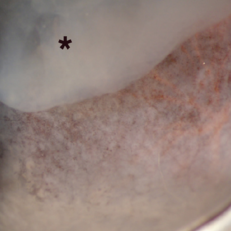 Toxoplasmic Acute Retinal Necrosis
May 18 2020 by McGill University Health Centre
Toxoplasmosis can have several manifestations in the eye, of which toxoplasmic acute retinal necrosis has the worst prognosis. This enucleation specimen shows extensive retinal necrosis with multiple coalescent foci. The vitreous is hazy (*).
Condition/keywords: acute retinal necrosis, toxoplasmosis
A project from the American Society of Retina Specialists