-
 Branch retinal artery occlusion
Jan 24 2023 by Rayna Marshall
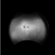
Widefield fundus autofluorescence image of a 54-year-old female with an asymptomatic chronic branch retinal artery occlusion in the left eye. Hyper-autofluorescent embolus present at proximal inferior arcade, hypo-autoflorescence temporally corresponding to hyper-pigmentation. Vision was 20/20.
Photographer: Drew H. Scoles, MD, PhD, University of Pennsylvania
Condition/keywords: branch retinal artery occlusion (BRAO), BRAO, embolus
-
 Branch retinal artery occlusion
Jan 24 2023 by Rayna Marshall
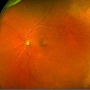
Widefield fundus image of a 54-year-old female with an asymptomatic chronic branch retinal artery occlusion in the left eye. Peripheral schisis-like changes with pigmentation and temporal dot-blot hemorrhages. Vision was 20/20.
Photographer: Drew H. Scoles, MD, PhD, University of Pennsylvania
Condition/keywords: branch retinal artery occlusion (BRAO), BRAO, embolus
-
 Branch retinal artery occlusion
Jan 24 2023 by Rayna Marshall
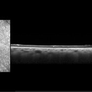
OCT image of a 54-year-old female with an asymptomatic chronic branch retinal artery occlusion in the left eye showing inner retinal atrophy in the inferior macula corresponding to the region of chronic ischemia. Vision was 20/20.
Photographer: Drew H. Scoles, MD, PhD, University of Pennsylvania
Condition/keywords: branch retinal artery occlusion (BRAO), BRAO, embolus
A project from the American Society of Retina Specialists