-
 Fluorescein Leakage from Choroidal Neovascularization Due to Age-Related Macular Degeneration in Absence of Abnormality on Spectral-Domain Optical Coherence Tomography
Feb 11 2013 by Neil M. Bressler, MD
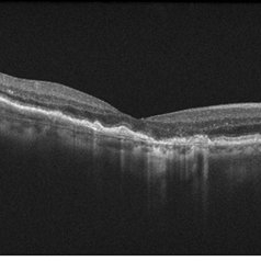
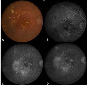
During monitoring of anti-vascular endothelial growth factor therapy in neovascular age-related macular degeneration (Fig A), 10% of active CNV with fluorescein angiographic leakage (Fig B - D) may show the absence of fluid in SD-OCT1 (Fig E), as shown in this case. Reference 1 Khurana RN, Dupas B, Bressler NM. Agreement of time-domain and spectral-domain optical coherence tomography with fluorescein leakage from choroidal neovascularization. Ophthalmology 2010;117:1376-1380.
Condition/keywords: age-related macular degeneration (AMD), choroidal neovascularization (CNV)
-
 Fluorescein Leakage from Choroidal Neovascularization Due to Age-Related Macular Degeneration in Absence of Abnormality on Spectral-Domain Optical Coherence Tomography
Feb 11 2013 by Neil M. Bressler, MD
During monitoring of anti-vascular endothelial growth factor therapy in neovascular age-related macular degeneration (Fig A), 10% of active CNV with fluorescein angiographic leakage (Fig B - D) may show the absence of fluid in SD-OCT1 (Fig E), as shown in this case. Reference 1 Khurana RN, Dupas B, Bressler NM. Agreement of time-domain and spectral-domain optical coherence tomography with fluorescein leakage from choroidal neovascularization. Ophthalmology 2010;117:1376-1380.
Condition/keywords: age-related macular degeneration (AMD), choroidal neovascularization (CNV)
-
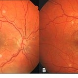
 Example of AREDS Category 1 (Small Drusen But Not Considered AMD)
Feb 11 2013 by Neil M. Bressler, MD
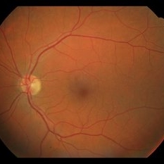
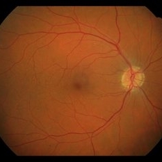
Person in AREDS Category 1 were essentially free of age-related macular abnormalities, with a total drusen area less than 5 small drusen (<63 microns) within 3,000 microns of the center of the macula, and visual acuity of 20/32 or better in both eyes1. These are fundus photographs of a 53-year-old man, with visual acuity 20/20 OD and 20/32 OS presenting for evaluation of any diabetic retinopathy. Reference: 1 Age-Related Eye Disease Study Research Group. A randomized, placebo controlled clinical trial of high-dose supplementation with vitamins C and E, beta carotene, and zinc for age-related macular degeneration and vision loss: AREDS report No. 8. Arch Ophthalmol. 2001;119(10):1417-1436.
Condition/keywords: age-related macular degeneration (AMD)
-
 Example of AREDS Category 1 (Small Drusen But Not Considered AMD)
Feb 11 2013 by Neil M. Bressler, MD
Person in AREDS Category 1 were essentially free of age-related macular abnormalities, with a total drusen area less than 5 small drusen (<63 microns) within 3,000 microns of the center of the macula, and visual acuity of 20/32 or better in both eyes1. These are fundus photographs of a 53-year-old man, with visual acuity 20/20 OD and 20/32 OS presenting for evaluation of any diabetic retinopathy. Reference: 1 Age-Related Eye Disease Study Research Group. A randomized, placebo controlled clinical trial of high-dose supplementation with vitamins C and E, beta carotene, and zinc for age-related macular degeneration and vision loss: AREDS report No. 8. Arch Ophthalmol. 2001;119(10):1417-1436.
Condition/keywords: age-related macular degeneration (AMD)
-
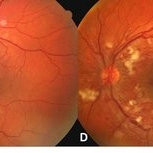 Worsening of Diabetic Retinopathy Severity Level Associated with Initiation of Tight Glucose Control
Feb 11 2013 by Neil M. Bressler, MD
Tight control of systemic blood sugar in juvenile-onset diabetes may lead to worsen diabetic retinopathy for a temporary period, and will lead to better retinopathy condition in longer term of the disease1. A 20-year-old man with Type 1 diabetes presented with severe non-proliferative diabetic retinopathy in both eyes at the initiation of intensive diabetes treatment (Fig A, C). 3 months after initiating tight blood sugar control, his fundus shows substantial worsening of this diabetic retinopathy severity level with new neovascularization of the disc, numerous new nerve fiber layer infarcts surrounding the optic nerve (in the absence of any systemic hypertension) and a marked increase in intraretinal hemorrhages in both eyes (Fig B, D). Reference: 1. The Diabetes Control and Complications Trial Research Group. The effect of intensive treatment of diabetes on the development and progression of long-term complications in insulin-dependent diabetes mellitus. N Engl J Med 1993; 329:977-986.
A project from the American Society of Retina Specialists