-
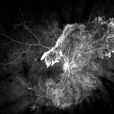 Proliferative Diabetic Retinopathy
Aug 22 2019 by Parnian Arjmand, MD, MSc, FRCSC, DABO, FASRS
Proliferative diabetic retinopathy.
Imaging device: Optos, IVFA
Condition/keywords: proliferative diabetic retinopathy (PDR)
-
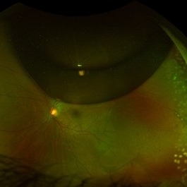 Chronic Retinal Detachment after Pneumatic Retinopexy
Jan 8 2022 by Parnian Arjmand, MD, MSc, FRCSC, DABO, FASRS
This is a fundus photo in the eye of a young phakic patent who presented with a 6 month history of "difficulty seeing at night" and subjective nasal "blurriness" in the left eye. There was a chronic temporal RD, OS, extending to the arcades (Mac on). This photo is week 1 s/p Pneumatic retinopexy with SF6 gas and laser retinopexy to temporal breaks (6 holes, lattice); no PVD. As you can see, there is a "bleb" of viscous schlieren given the chronic nature of this RD that persist posterior to the breaks and temporal to the macula. This type of sub retinal fluid may take months to years to resorb.
Condition/keywords: chronic retinal detachment, pneumatic retinopexy
-
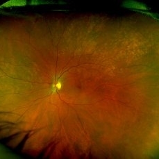 Rubella retinopathy
Jan 8 2022 by Parnian Arjmand, MD, MSc, FRCSC, DABO, FASRS
Bilateral pigmentary retinopathy in a young patient with a history of congenital rubella.
Condition/keywords: bilateral pigmentary retinopathy, congenital rubella, rubella
-
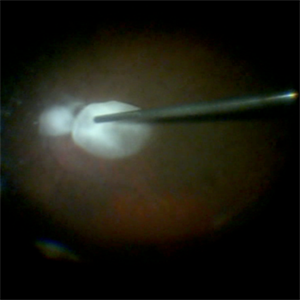 Staining-the-vitreous
Jan 10 2022 by Parnian Arjmand, MD, MSc, FRCSC, DABO, FASRS
This is an example of staining the posterior hyaloid in an eye with no PVD with triamcinolone actinide to assist with inducing a PVD prior to repairing the macular hole.
Condition/keywords: macular hole, PVD, triamcinolone, triescence
-
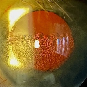 PFO Bubbles
Feb 25 2025 by Parnian Arjmand, MD, MSc, FRCSC, DABO, FASRS
Post operative day 7 after repair of an RD secondary to a giant retinal tear with temporary PFO tamponade.
Condition/keywords: GRT, PFO
-
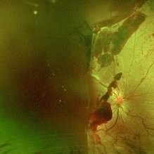 PDR
PDR
Feb 25 2025 by Parnian Arjmand, MD, MSc, FRCSC, DABO, FASRS
Young patient with proliferative diabetic retinopathy and subhyaloid hemorrhage. Extensive neovascular fronds can be noted throughout the posterior pole and disc.
Condition/keywords: subhyaloid hemorrhage
-
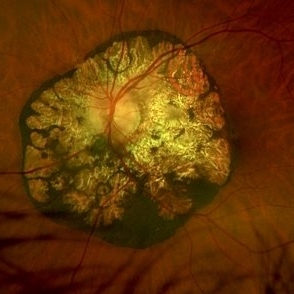 Peripapillary CHRPE
Aug 29 2025 by Parnian Arjmand, MD, MSc, FRCSC, DABO, FASRS
Optos color photo of a 64-year old male with an incidental finding of peri papillary congenital hypertrophy of the retinal pigment epithelium.
Condition/keywords: Peripapillary CHRPE
-
 Dislocated Lens
Dec 8 2025 by Parnian Arjmand, MD, MSc, FRCSC, DABO, FASRS
A high myope patient presented 12 years after PPV a Cataract extraction for a retinal detachment repair with a new onset of vision loss. A dislocated IOL was noted on clinical examination.
Condition/keywords: Aphakia, dropped IOL, myopia, zonular dehiscence
-
 CSNB-OS
Feb 25 2026 by Parnian Arjmand, MD, MSc, FRCSC, DABO, FASRS
A middle-aged female presented with long-standing nyctalopia and excellent visual acuity. She reported no known family history of inherited eye disease. Dilated fundus examination demonstrated multiple, discrete, yellow-white punctate lesions throughout the posterior pole and mid-periphery, consistent with fundus albipunctatus within the congenital stationary night blindness (CSNB) spectrum. The optic nerves appeared healthy without pallor, and the retinal vasculature showed no attenuation. Left eye shown above.
Condition/keywords: fundus albipunctatus
-
 CSNB-OD
Feb 25 2026 by Parnian Arjmand, MD, MSc, FRCSC, DABO, FASRS
A middle-aged female presented with long-standing nyctalopia and excellent visual acuity. She reported no known family history of inherited eye disease. Dilated fundus examination demonstrated multiple, discrete, yellow-white punctate lesions throughout the posterior pole and mid-periphery, consistent with fundus albipunctatus within the congenital stationary night blindness (CSNB) spectrum. The optic nerves appeared healthy without pallor, and the retinal vasculature showed no attenuation. Right eye shown above.
Imaging device: Optos California
Condition/keywords: congenital stationary night blindness (CSNB), fundus albipunctatus
-
 Dislocaed IOL in Retinitis Pigmentosa
Oct 15 2026 by Parnian Arjmand, MD, MSc, FRCSC, DABO, FASRS
A lone one piece acrylic IOL sits on an RP retina in the eye of a patient with an optically empty vitreoretinopathy
Photographer: Alex Amigleo
Condition/keywords: aphakia, sector retinitis pigmentosa

A project from the American Society of Retina Specialists