-
 Secondary CNVM
Apr 21 2022 by Maneesh M Bapaye, MD, MBA
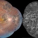
The image depicts montage color photograph as well as montage fluorescine angiography image of healed multifocal choroiditis with seconadory subfoveal cnvm and subretinal haemorrhage in a 28 years old female patient
Photographer: Maneesh Bapaye, Dr.Bapaye Hospital, Nashik, India
Condition/keywords: healed choroiditis, seconadry cnvm
-
 Spent-force
Jun 27 2023 by Maneesh M Bapaye, MD, MBA
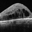
Composite OCT image of left eye of a 53 years old male diabetic patient with recuuent spongy diabetic macular edema. Structural image depicts presence of ozurdex temporal to fovea. B-Scan image shows localized action of ozurdex in retinal tissue underlying it while cystoid changes are seen nasal to foveal center
Photographer: Maneesh Bapaye MD
Condition/keywords: diabetic macular edema, Ozurdex implant
-
 CRAO with cilioretinal sparing - Multimodal imaging
Jun 28 2023 by Maneesh M Bapaye, MD, MBA
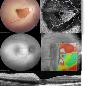
A 34 year old male patient presented with sudden onset vision loss of 1 week duration. Visual acuity at presentation was 20/200. Fundus examination revealed diffuse retinal whitening with sparing of papillomacular bundle and fovea due to patent cilioretinal artery. Autofluorescence shows peripheral hypoAF, patent capillaries can be seen only in area of cilioretinal supply, OCT shows thickening of inner retinal layers temporal to fovea Systemic examination revealed that patient had valvular heart disease with multiple valves involved.
Photographer: Maneesh Bapaye
Condition/keywords: cilioretinal sparing, CRAO, multimodal imaging
-
 Iridodialysis Repair
Jul 11 2023 by Maneesh M Bapaye, MD, MBA
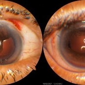
A 68 year old female patient was referred with large iridodialysis in superotemporal and temporal quadrant in right eye, as seen in left side image in the panel. She underwent iridodialysis repair using 9-0 prolene suture on single armed needle to suture the iris to iris root in hangback fashion. The patient had satisfactory anatomical outcome as seen in right side image in the panel.
Photographer: Dr.Maneesh Bapaye
Condition/keywords: Intraoperative iridodialysis
-
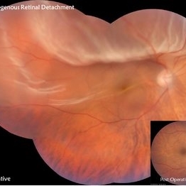 Retinal Detachment
Jul 30 2023 by Maneesh M Bapaye, MD, MBA
Montage fundus photo of superior retinal detachment in right eye of a 45 year old male patient
Photographer: Dr.Maneesh Bapaye
Imaging device: Zeiss Fundus camera
Condition/keywords: montage fundus photo
-
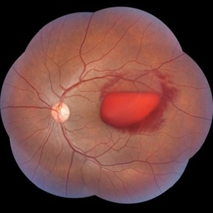 Subhyaloid Haemorrhage - Valsalva Retinopathy
Jul 30 2023 by Maneesh M Bapaye, MD, MBA
A 25 years old paitient presented with sudden loss of vision following sudden rise intra abdominal pressure, Montage photo
Photographer: Dr.Maneesh Bapaye
Imaging device: Zeiss Fundus Camera
Condition/keywords: subhyaloid hemorrhage, valsalva retinopathy
-
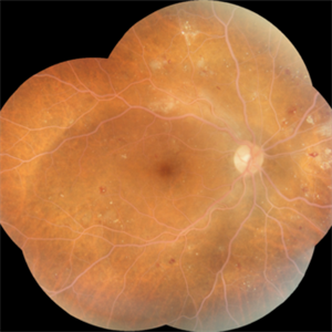 Lipemia Retinalis -Right eye
Aug 1 2023 by Maneesh M Bapaye, MD, MBA
A 52 years old poorly controlled diabetic male pt. HbA1c of 16.2%, Sr. Cholesterol 460, Sr. Triglycerides 575, VLDL 115
Photographer: Dr.Maneesh Bapaye
Imaging device: Zeiss fundus camera
Condition/keywords: clinically significant macular edema (CSME), lipemia retinalis, nonproliferative diabetic retinopathy
-
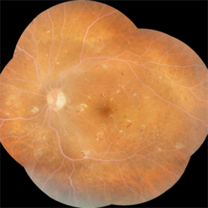 Lipemia retinalis with severe NPDR- Left Eye
Aug 1 2023 by Maneesh M Bapaye, MD, MBA
A 52 years old poorly controlled diabetic male pt. HbA1c of 16.2%, Sr. Cholesterol 460, Sr. Triglycerides 575, VLDL 115
Photographer: Dr.Maneesh Bapaye
Imaging device: Zeiss fundus camera
Condition/keywords: clinically significant macular edema (CSME), lipemia retinalis, nonproliferative diabetic retinopathy
A project from the American Society of Retina Specialists