-
 Retinoschisis with Retinal detachment
Jan 3 2023 by Shobhit Chawla, M.S.
Retinoschisis with Retinal detachment
Photographer: Shobhit Chawla
Imaging device: ZeissClarus500
Condition/keywords: RETINOSCHISIS
-
 Retinoschisis with Retinal detachment
Jan 3 2023 by Shobhit Chawla, M.S.
Retinoschisis with Retinal detachment
Photographer: Shobhit Chawla
Imaging device: ZeissClarus500
Condition/keywords: RETINOSCHISIS
-
 Idiopathic Vasculitis
Aug 29 2023 by Shobhit Chawla, M.S.
A 28 year old male with acute loss of vision and no history of any illness. Had profound loss of vision with severe vasulitis extending from central fundus to periphery.
Photographer: Ranjit Ray
Imaging device: Clarus
Condition/keywords: VASCULITIS
-
 Idiopathic vasculitis
Aug 29 2023 by Shobhit Chawla, M.S.
A 28 year old male with acute loss of vision and no history of any illness. Had profound loss of vision with severe vasulitis extending from central fundus to periphery.
Photographer: Ranjit Ray
Imaging device: Clarus
Condition/keywords: VASCULITIS
-
 Traumatic Macular Hole pre and post repair
Nov 25 2024 by Shobhit Chawla, M.S.
31 year-old male reported with h/o of blunt trauma over right eye ,from cricket ball. On examination DVA RE 6/60,LE 6/18,ant segment BE :WNL,FUNDUS RE:Sub retinal hemorrhage at macula with chroidal tear,LE :WNL. Undwer went 25G vitrectomy+sub retinal TPA+C3F8(RE).Post op 1 month DVA RE:6/24 ,ANT SEGMENT:WNL,FUNDUS:resolved sub retinal haem with traumatic macular hole. Under went repeat vit+autologous retinal transplant +SOI RE.POST SOR AFTER4monthsV/A :6/18 RE
Photographer: Ranjit Ray
Imaging device: Clarus 500
Condition/keywords: Macular hole, retinal graft, subretinal hemorrhage
-
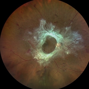 Table Top TRD
Dec 5 2024 by Shobhit Chawla, M.S.
50 year old diabetic male with table top TRD and fair visual acuity .
Photographer: Ranjit Ray
Imaging device: Clarus500
Condition/keywords: florid type PDR, PDR, TRD
-
 Proliferative Diabetic Retinopathy
Dec 5 2022 by Shobhit Chawla, M.S.
36 year old gentleman , with IDDM and severe high risk PDR. Pre and post pattern scan PRP. This image pre PRP.
Photographer: Shobhit Chawla
Imaging device: ZeissClarus500
Condition/keywords: HRC-PDR, IDDM, proliferative diabetic retinopathy (PDR)
-
 Proliferative Diabetic Retinopathy
Dec 5 2022 by Shobhit Chawla, M.S.
36 year old gentleman , with IDDM and severe high risk PDR. Pre and post pattern scan PRP. This image post PRP
Photographer: Shobhit Chawla
Imaging device: ZeissClarus500
Condition/keywords: HRC-PDR, proliferative diabetic retinopathy (PDR)
-
 PVD induction with IVTA staining
Nov 1 2022 by Shobhit Chawla, M.S.
This is an intraoperative photograph of Pvd induction showing both the macular and disc attatchments stained with triamcinolone.
Photographer: Shobhit Chawla
Condition/keywords: PVD induction, triamcinolone
-
 Peripheral haemorrhagic. Choroidopathy
May 12 2022 by Shobhit Chawla, M.S.
An elderly with peripheral subretinal haemorrhage due to a peripheral haemorrhagic choroidopathy akin to a peripheral PCV like lesion
Photographer: Shobhit chawla
Imaging device: Zeiss clarus 500
Condition/keywords: PERIPHERAL HAEMORRHAGIC CHOROIDOPATHY
-
 Submacular haemorrhage PCV
May 6 2022 by Shobhit Chawla, M.S.
SUBMACULAR HAEMORRHAGE IN A 38YEAR OLD LADY PATIENT CAUSE POLYP BLEED IN PCV. Following viterectomy , subretinal tpa . gas and aflibercept injection. 1ST day post operative image.
Photographer: Shobhit Chawla
Imaging device: ZeissClarus500
Condition/keywords: aflibercept, idiopathic polypoidal choroidal vasculopathy, polypoidal choroidal vasculopathy (PCV), tissue plasminogen activator (tPA)
-
 Post Subretinal tpa , viterectomy and gas
May 6 2022 by Shobhit Chawla, M.S.
SUBMACULAR HAEMORRHAGE IN A 38YEAR OLD LADY PATIENT CAUSE POLYP BLEED IN PCV. Following viterectomy , subretinal tpa . gas and aflibercept injection. 7 day post operative image.
Photographer: Shobhit Chawla
Imaging device: Zeiss Clarus 500
Condition/keywords: aflibercept, intravitreal gas bubble, submacular hemorrhage, tissue plasminogen activator (tPA), vitrectomy
-
 PCV and Peripheral Hemorrhagic Choroidopathy
Feb 25 2022 by Shobhit Chawla, M.S.
PCV and Peripheral Hemorrhagic Choroidopathy in right eye.
Photographer: Shobhit Chawla
Imaging device: Zeiss Clarus 500
Condition/keywords: PERIPHERAL HAEMORRHAGIC CHOROIDOPATHY, polypoidal choroidal vasculopathy (PCV)
-
 Branching vascular network..Polypoidal choroidal vasculopathy
Feb 21 2022 by Shobhit Chawla, M.S.
A 56 year old patient on treatment with ANTIVGEF therapy for six years
Photographer: Shobhit Chawla
Imaging device: Zeiss Clarus 500
Condition/keywords: branching vascular network (BVN), polypoidal choroidal vasculopathy (PCV)
A project from the American Society of Retina Specialists