-
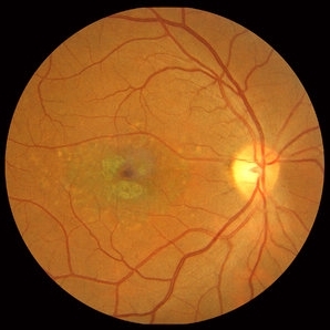 Stargardt macular dystrophy slide 1
Oct 22 2012 by Ronald C. Gentile, MD
16-year-boy with difficulty in school seeing the black board. The macula area of the right eye had areas with a beaten bronze appearance and atrophy. Small pisci-form flecks can be seen surrounding the fovea.
Photographer: The New York Eye & Ear Infirmary Department of Medical Imaging
Condition/keywords: small pisci-form flecks, Stargardt disease
-
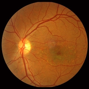 Stargardt macular dystrophy slide 2
Oct 22 2012 by Ronald C. Gentile, MD
Fundus examination of the left eye had similar findings with centrally atrophic macula area with surrounding flecks.
Photographer: The New York Eye & Ear Infirmary Department of Medical Imaging
Condition/keywords: Stargardt disease
A project from the American Society of Retina Specialists