-
 Serpiginous Choroidal Atrophy
Mar 29 2019 by Jessica Norkus
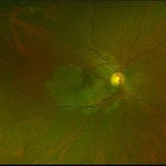
Optos ultra wide field color image of 20-year-old female presenting with serpiginous choroidal atrophy. Patient was unaware of vision loss OD, until accidentally covering OS and noticing the change. Acuity of 20/200 OD and 20/15 OS at time of imaging.
Photographer: Jessica Norkus
Imaging device: Optos Wide Field Camera
Condition/keywords: abnormal fundus, color fundus photograph, fundus photograph, macula serpiginous choroidopathy, Optomap, Optos, ultra-wide field imaging
-
 Serpiginous Choroidal Atrophy
Mar 29 2019 by Jessica Norkus
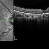
Heidelberg single horizontal scan image of 20-year-old female presenting with serpiginous choroidal atrophy. Patient was unaware of vision loss OD, until accidentally covering OS and noticing the change. Acuity of 20/200 OD and 20/15 OS at time of imaging.
Photographer: Jessica Norkus
Imaging device: Heidelberg Spectralis
Condition/keywords: Heidelburg Spectralis, macula lesion, macula serpiginous choroidopathy, optical coherence tomography (OCT)
-
 Serpiginous Choroidal Atrophy
Mar 29 2019 by Jessica Norkus
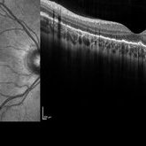
Heidelberg single vertical scan image of 20-year-old female presenting with serpiginous choroidal atrophy. Patient was unaware of vision loss OD, until accidentally covering OS and noticing the change. Acuity of 20/200 OD and 20/15 OS at time of imaging.
Photographer: Jessica Norkus
Imaging device: Heidelberg Spectralis
Condition/keywords: Heidelburg Spectralis, macula lesion, macula serpiginous choroidopathy, optical coherence tomography (OCT)
-
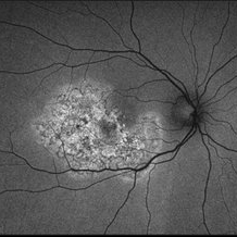
 Serpiginous Choroidal Atrophy
Mar 29 2019 by Jessica Norkus
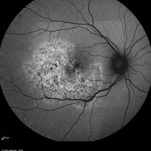
50 degree Auto fluorescent image of 20-year-old female presenting with serpiginous choroidal atrophy. Patient was unaware of vision loss OD, until accidentally covering OS and noticing the change. Acuity of 20/200 OD and 20/15 OS at time of imaging.
Photographer: Jessica Norkus
Imaging device: Heidelberg Spectralis
Condition/keywords: Heidelburg Spectralis, macula lesion, macula serpiginous choroidopathy, optical coherence tomography (OCT), wide angle imaging
-
 Serpiginous Choroidal Atrophy
Mar 29 2019 by Jessica Norkus
Optos ultra wide field auto fluorescent image of 20-year-old female presenting with serpiginous choroidal atrophy. Patient was unaware of vision loss OD, until accidentally covering OS and noticing the change. Acuity of 20/200 OD and 20/15 OS at time of imaging.
Photographer: Jessica Norkus
Imaging device: Optos Ultra Wide Field Camera
Condition/keywords: fundus autofluorescence (FAF), fundus photograph, macula lesion, macula serpiginous choroidopathy, Optos, ultra-wide field imaging
A project from the American Society of Retina Specialists