-
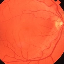
 Central Serous Retinopathy
Mar 26 2019 by Gary R. Cook, MD, FACS
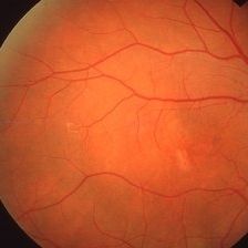
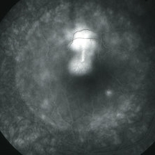
45-year-old white male with history of bilateral CSR; Color fundus photo showing an area of RPE depigmentation in temporal macula; VA = 20/200.
Imaging device: Topcon VT-50
Condition/keywords: central serous retinopathy (CSR)
-
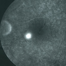
 Central Serous Retinopathy
Mar 26 2019 by Gary R. Cook, MD, FACS
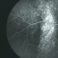
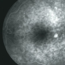
Mid-phase FA frame of right eye of a 45-year-old white male with a history of bilateral CSR showing stippled hyperfluorescence in an area of an RPE track beneath the inferotemporal arcade OD; no active dye leakage is present; VA = 20/200.
Imaging device: Topcon VT-50
Condition/keywords: central serous retinopathy (CSR), FA mid phase
-
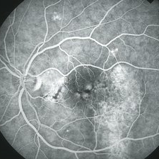
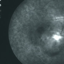
 Central Serous Retinopathy
Mar 26 2019 by Gary R. Cook, MD, FACS
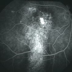
Late-phase FA frame of the right eye of a 45-year-old white male with a history of CSR showing pooling of dye beneath a small chronic RPED near fovea and stippled late staining of the RPE track in macula and beneath the inferotemporal arcade OD; VA = 20/200.
Imaging device: Topcon VT-50
Condition/keywords: central serous retinopathy (CSR), FA late phase
-
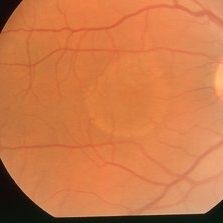
 Central Serous Retinopathy
Mar 26 2019 by Gary R. Cook, MD, FACS
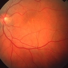
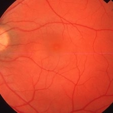
45-year-old white male with a history of bilateral CSR showing subtle RPE depigmentation in macula and inferiorly in his symptomatic OS; VA = 20/30.
Imaging device: Topcon VT-50
Condition/keywords: central serous retinopathy (CSR)
-
 Central Serous Retinopathy
Mar 26 2019 by Gary R. Cook, MD, FACS
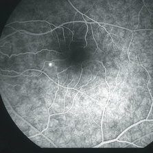
Venous phase of FA of left eye of 45-year-old white male with CSR showing multiple RPE abnormalities and a depigmented RPE track in temporal macula going inferiorly; VA= 20/30.
Imaging device: Topcon VT-50
Condition/keywords: central serous retinopathy (CSR), FA mid phase
-
 Central Serous Retinopathy
Mar 26 2019 by Gary R. Cook, MD, FACS
Late-phase frame of FA of 45-year-old white male with CSR OS showing stippled staining in area of RPE track temporally and pooling of dye beneath 3 - 4 small RPEDs OS; VA = 20/30.
Imaging device: Topcon VT-50
Condition/keywords: central serous retinopathy (CSR), FA late phase, staining
-
 Bilateral Central Serous Retinopathy
Mar 26 2019 by Gary R. Cook, MD, FACS
Right eye of a 37-year-old white male with a history of bilateral CSR showing a 2 DD NSRD centrally in his symptomatic OD; VA = 20/20-2.
Imaging device: Topcon VT-50
Condition/keywords: central serous retinopathy (CSR), neurosensory detachment of retina
-
 Bilateral Central Serous Retinopathy
Mar 26 2019 by Gary R. Cook, MD, FACS
Late-phase fluorescein angiogram image of the right eye of a 37-year-old white male showing pinpoint leak with late diffusion of dye from it superiorly and RPE irregularities nasal to fovea in a case of bilateral central serous retinopathy; VA = 20/20-2.
Imaging device: Topcon VT-50
Condition/keywords: central serous retinopathy (CSR), FA late phase
-
 Bilateral Central Serous Retinopathy
Mar 26 2019 by Gary R. Cook, MD, FACS
Late-phase frame of FA of 37-year-old white male with acute CSR OD showing pooling of dye beneath the small central RPED centrally, a smokestack-type leak from the RPE defect just above it, and mild late pooling of dye outlining the large neurosensory macular detachment; VA = 20/80-1.
Imaging device: Topcon VT-50
Condition/keywords: central serous retinopathy (CSR), FA late phase, FA late phase leakage, neurosensory detachment of retina
-
 Bilateral Central Serous Retinopathy
Mar 26 2019 by Gary R. Cook, MD, FACS
Asymptomatic left eye of a 37-year-old white male with a history of previous CSR OS showing some focal RPE depigmentation perifoveally and subretinic deposits temporally; no NSRD is present; VA = 20/15+3.
Imaging device: Topcon VT-50
Condition/keywords: central serous retinopathy (CSR), resolved subretinal fluid, retinal pigment epithelium (RPE) changes
-
 Bilateral Central Serous Retinopathy
Mar 26 2019 by Gary R. Cook, MD, FACS
Mid-phase fluorescein angiogram frame of a pinpoint leak in the temporal macula OS of a 37-year-old white male with bilateral central serous retinopathy; VA = 20/15+3.
Imaging device: Topcon VT-50
Condition/keywords: central serous retinopathy (CSR), FA mid phase
-
 Central Serous Retinopathy
Mar 26 2019 by Gary R. Cook, MD, FACS
32-year-old white female with acute CSR with 1.5DD serous detachment in the macula OS; VA = 20/30-2.
Imaging device: Topcon VT-50
Condition/keywords: central serous retinopathy (CSR), neurosensory detachment of retina
-
 Central Serous Retinopathy
Mar 26 2019 by Gary R. Cook, MD, FACS
32 year old white female with acute CSR OS; early venous filling phase of FA showing a pinpoint leak in inferonasal macula; VA = 20/30-2.
Imaging device: Topcon VT-50
Condition/keywords: central serous retinopathy (CSR), FA mid phase
-
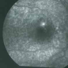
 Central Serous Retinopathy
Mar 26 2019 by Gary R. Cook, MD, FACS
32-year-old white female with acute CSR OS; 10-minute late frame of FA OS showing diffusion of dye from earlier pinpoint leak in inferonasal macula; VA = 20/30-2.
Imaging device: Topcon VT-50
Condition/keywords: central serous retinopathy (CSR), FA late phase leakage
-
 CSR with RPED
Mar 26 2019 by Gary R. Cook, MD, FACS
White male with acute CSR with a RPED beneath the NSRD OD.
Imaging device: Topcon VT-50
Condition/keywords: central serous retinopathy (CSR), retinal pigment epithelium (RPE) detachment
-
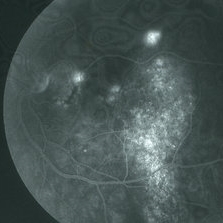
 CSR with large RPED
Mar 26 2019 by Gary R. Cook, MD, FACS
Mid-phase FA showing large RPED inferonasal to optic disc with overlying cruciate pigment figures (black lines) and neurosensory macular detachment OD.
Imaging device: Topcon VT-50
Condition/keywords: central serous retinopathy (CSR), neurosensory detachment of retina, retinal pigment epithelium (RPE) detachment
-
 CSR with large RPED
Mar 26 2019 by Gary R. Cook, MD, FACS
Late-phase FA frame showing mild pooling of dye beneath a large RPED inferonasal to the optic disc, blocked fluorescence from the pigment figures (black lines), and late dye leakage from the RPED.
Imaging device: Topcon VT-50
Condition/keywords: central serous retinopathy (CSR), FA late phase, retinal pigment epithelium (RPE) detachment
-
 Central Serous Retinopathy
Mar 26 2019 by Gary R. Cook, MD, FACS
43-year-old white male with acute CSR OD showing a large neurosensory macular detachment in his right eye.
Imaging device: Topcon VT-50
Condition/keywords: central serous retinopathy (CSR), neurosensory detachment of retina
-
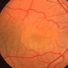
 Chronic CSR
Mar 26 2019 by Gary R. Cook, MD, FACS
46-year-old male with chronic (>6 months duration) CSR OD; VA= 20/30-2.
Imaging device: Topcon VT-50
Condition/keywords: chronic central serous chorioretinopathy (CSCR)
A project from the American Society of Retina Specialists