-
 Slide 3-1
Feb 19 2019 by Lancaster Course in Ophthalmology
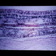
Low- power view of sclera, choroid, and retinal pigment epithelium, showing diffuse chronic granulomatous inflammation in sympathetic ophthalmia ( x25).
Condition/keywords: choroid, chronic granulomatous inflammation, ophthalmia, retinal pigment epithelium, sclera
-
 Slide 3-2
Feb 19 2019 by Lancaster Course in Ophthalmology
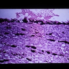
Higher-power view of the same sympathetic ophthalmic eye, showing giant cells and epithelioid cells (x65).
Condition/keywords: epithelioid cells, ophthalmia
-
 Slide 3-3
Feb 19 2019 by Lancaster Course in Ophthalmology
Cluster of epithelioid cells in choroid of eye with sympathetic ophthalmia (x160).
Condition/keywords: choroid, epithelioid cells, ophthalmia
-
 Slide 3-4
Feb 19 2019 by Lancaster Course in Ophthalmology
Serous detachment of retina overlying area of infiltrated choroid in sympathetic ophthalmia (x12).
Condition/keywords: choroid, serous retinal detachment
-
 Slide 3-5
Feb 20 2019 by Lancaster Course in Ophthalmology
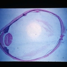
Low-power view showing entire eye in end stage of phacoanaphylactic endophthalmitis. Note that the lens is the central focus of pathology and is extensively resorbed. Note also the occlusion and seclusion of the pupil as well as the cyclitic membrane and total retinal detachment.
Condition/keywords: cyclitic membrane, endophthalmitis
-
 Slide 3-6
Feb 20 2019 by Lancaster Course in Ophthalmology
Higher-power view of lens area in eye with phacoanaphylaxis ( x120). Note the zonular type granulomatous reaction and adhesion of iris.
Condition/keywords: granulomatous reaction, iris, phacoanaphylaxis
-
 Slide 3-7
Feb 20 2019 by Lancaster Course in Ophthalmology
Phacoanaphylaxis showing residual lens material, granulomatous inflammation, and granulation tissue ( x25).
Condition/keywords: granulation tissue, phacoanaphylaxis
-
 Slide 3-8
Feb 20 2019 by Lancaster Course in Ophthalmology
Destruction of choroid and retina in endophthalmitis caused by Nocardia asteroides, a bacilliform, gram-positive, acid-fast bacillus ( x65).
Condition/keywords: bacilliform, choroid, endophthalmitis, Nocardia asteroides
-
 Slide 3-9
Feb 20 2019 by Lancaster Course in Ophthalmology
Low-power view of eye with Candida albicans endophthalmitis ( x12). Note the hypopyon and exudate in the anterior chamber and the infiltration of the iris and ciliary body with inflammatory cells.
Condition/keywords: ciliary, endophthalmitis, exudate, hypopyon, iris
-
 Slide 3-10
Feb 20 2019 by Lancaster Course in Ophthalmology
Candida albicans involving the cornea ( x65).
Condition/keywords: cornea, endophthalmitis
-
 Slide 3-11
Feb 20 2019 by Lancaster Course in Ophthalmology
Higher-power view of hyphae and spores of Candida albicans in Candida keratitis ( x160).
Condition/keywords: endophthalmitis, hyphae, keratitis, spores
-
 Slide 3-12
Feb 20 2019 by Lancaster Course in Ophthalmology
Branching nonseptate fungi seen in ocular infection with mucormycosis ( x160).
Condition/keywords: infection, mucormycosis, non septate fungi
-
 Slide 3-13
Feb 20 2019 by Lancaster Course in Ophthalmology
Cystoid macular change with granulomatous inflammation of choroid and retina in eye of patient with presumed histoplasmosis retinochoroiditis ( x25).
Condition/keywords: choroid, cystoid macular degeneration, granulomatous inflammation, histoplasmosis
-
 Slide 3-14
Feb 20 2019 by Lancaster Course in Ophthalmology
Adjacent area to that in Slide 3-13, showing degenerative change and inflammatory infiltrate in presumed histoplasmosis retinochoroiditis ( x25).
Condition/keywords: histoplasmosis, inflammatory infiltrate
-
 Slide 3-15
Feb 20 2019 by Lancaster Course in Ophthalmology
Higher-power view of choroid in presumed histoplasmosis retinochoroiditis, showing marked epithelioid cell infiltrate ( x65).
Condition/keywords: choroid
-
 Slide 3-16
Feb 20 2019 by Lancaster Course in Ophthalmology
Enucleated eye with Toxocara endophthalmitis, showing severe posterior inflammation with retinal detachment and disorganization.
Condition/keywords: endophthalmitis, enucleation, toxocariasis
-
 Slide 3-18
Feb 20 2019 by Lancaster Course in Ophthalmology
Area of anterior segments of eye with Toxocara endophthalmitis, showing iris infiltration by inflammatory cells, ectropion uveae, and angle closed by anterior synechiae ( x12).
Condition/keywords: anterior synechiae, ectropion uveae, endophthalmitis, iris, toxocariasis
-
 Slide 3-19
Feb 20 2019 by Lancaster Course in Ophthalmology
Low-power view of eye enucleated with sarcoid uveitis. Note the thickening of the choroid and iris.
Condition/keywords: choroid, iris, sarcoid
-
 Slid 3-20
Feb 20 2019 by Lancaster Course in Ophthalmology
View of iris and ciliary body in eye with sarcoid uveitis ( x12). Note the inflammatory cell infiltration of these structures.
Condition/keywords: ciliary, iris, sarcoid
-
 Slide 3-21
Feb 20 2019 by Lancaster Course in Ophthalmology
Higher-power view of ciliary body of eye shown in Slides 3-19 and 3-20 reveals presence of epithelioid and giant cells ( x65).
Condition/keywords: ciliary, epithelioid cells, giant cell
-
 Slide 3-22
Feb 20 2019 by Lancaster Course in Ophthalmology
View of choroid in eye with sarcoid uveitis, lymphocytes, and epithelioid cells with extension to sclera and degenerative changes in retinal pigment epithelium ( x65) .
Condition/keywords: choroid, epithelioid cells, epithelium, lymphocytes, sarcoid, sclera
-
 Slide 3-23
Feb 20 2019 by Lancaster Course in Ophthalmology
Conjunctival nodule removed from lower fornix of patient with sarcoid. A discrete pattern of granulomatous inflammation is evident ( x12).
Condition/keywords: conjunctival nodule, granulomatous inflammation, lower fornix, sarcoid
-
 Slide 3-24
Feb 20 2019 by Lancaster Course in Ophthalmology
Higher-power view showing noncaseating tubercle with Langhans' giant cells ( x65).
Condition/keywords: Langhans giant cell, noncaseating tubercle
-
 Slide 3-25
Feb 20 2019 by Lancaster Course in Ophthalmology
Lacrimal gland involved with granulomatous inflammation in patient with sarcoid. Note the discrete pattern.
Condition/keywords: granulomatous inflammation, lacrimal glands, sarcoid
-
 Slide 3-26
Feb 20 2019 by Lancaster Course in Ophthalmology
Higher-power view of Slide 3-25, showing epithelioid and giant cells admixed with epithelioid and plasma cells ( x25).
Condition/keywords: epithelioid cells, giant cell, plasma cells
-
 Slide 3-27
Feb 20 2019 by Lancaster Course in Ophthalmology
Low-power view of chalazion. Note the proximity of the inflammation to the meibomian glands.
Condition/keywords: chalazion, inflammation, meibomian glands
-
 Slide 3-28
Feb 20 2019 by Lancaster Course in Ophthalmology
Area of chalazion showing lipid droplets represented by empty spaces and predominantly chronic inflammation.
Condition/keywords: chalazion, chronic inflammation, lipid
-
 Slide 3-29
Feb 20 2019 by Lancaster Course in Ophthalmology
Pyogenic granuloma which frequently follows chalazion ( x65). In spite of its name it is neither pyogenic nor a granuloma. Note the profusion of newly formed blood vessels, inflammatory cells, and connective tissue.
Condition/keywords: chalazion, granuloma
-
 Slide 3-30
Feb 20 2019 by Lancaster Course in Ophthalmology
Low-power view of eye with Vogt-Koyanagi-Harada disease. Note the uveal infiltration.
Condition/keywords: uveal infiltration, Vogt-Koyanagi-Harada
-
 Slide 3-31
Feb 20 2019 by Lancaster Course in Ophthalmology
Higher-power view of choroid in Vogt-Koyanagi-Harada disease ( x65). Note the infiltration of the choroid. In contrast to sympathetic ophthalmia, there is no sparing of the choriocapillaris, and there is destruction of the retinal pigment epithelium.
Condition/keywords: choriocapillaris, choroid, ophthalmia, retinal pigment epithelium, Vogt-Koyanagi-Harada
A project from the American Society of Retina Specialists