69 yo WF found to have a "supero-nasal darkened elevation" in the left eye, during cataract evaluation. Patient was referred to Dr. Hruby, who confirmed that the elevation was a varix of the vortex vein; photos were obtained and thought to be good to put on Retina Image Bank. On the optos camera, the area of elevation was not seen in primary gaze. There is one pic with patient looking immediately supero-nasal (no varix seen) and another photo when patient holding a supero-nasal gaze (varix becomes present)
-
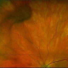 Varix of Vortex Vein - Optos Photo - Holding Superonasal Gaze
Jan 29 2019 by John S. King, MD
69-year-old white female found to have a "supero-nasal darkened elevation" in the left eye, during cataract evaluation. Patient was referred to Dr. Hruby, who confirmed that the elevation was a varix of the vortex vein; photos were obtained and thought to be good to put on Retina Image Bank. On the optos camera, the area of elevation was not seen in primary gaze. This is a picture of patient holding a gaze up and nasal. Varix seen supero-nasal.
Photographer: Karin Aletter
Imaging device: Optos California
Condition/keywords: vortex vein, vortex vein varix
-
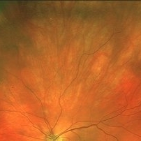 Varix of Vortex Vein - Optos, Immediately Looking Supero-Nasally
Jan 29 2019 by John S. King, MD
69-year-old white female found to have a "supero-nasal darkened elevation" in the left eye, during cataract evaluation. Patient was referred to Dr. Hruby, who confirmed that the elevation was a varix of the vortex vein; photos were obtained and thought to be good to put on Retina Image Bank. On the optos camera, the area of elevation was not seen in primary gaze. This is a picture of patient looking immediately up and nasal. Varix not seen.
Photographer: Karin Aletter
Imaging device: Optos California
Condition/keywords: vortex vein, vortex vein varix