-
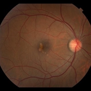 Unilateral PIC Following Recent Influenza Vaccine
Jan 6 2019 by John S. King, MD
42-year-old African American female with high myopia and type 2 diabetes, presented to her eye doctor with distortion in the right eye "that looked like seeing through a Coke bottle." She denied any photopsias or other symptoms. She received an influenza vaccine two weeks before onset of metamorphopsia. I saw her about a month after symptoms began. Va cc 20/30 OD J1 (20/15 J1+ OS); a/c and vitreous without cell or flare. Posterior pole OD showed yellowish, rounded small to medium RPE pigment alterations without heme or exudes (OS U/R). FA showed early focal areas of hyperfluorescence that stained in the later frames without CNVM or CME; rare MA inferiorly. The OCT showed some focal PEDs with some possible overlying SRHRM. We discussed options and decided to try a medrol dose pack. A few weeks later she was 20/20 J1, with minimal to no symptoms; OCT shows near total resolution of PEDs.
Photographer: Kay Dalby
Imaging device: Topcon 50
Condition/keywords: punctate inner choroidopathy (PIC), white dot syndrome
-
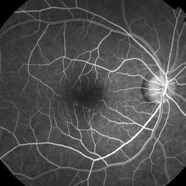 Unilateral PIC following recent influenza vaccine
Jan 6 2019 by John S. King, MD
FA 22 seconds 42-year-old African American female with high myopia and type 2 diabetes, presented to her eye doctor with distortion in the right eye "that looked like seeing through a Coke bottle." She denied any photopsias or other symptoms. She received an influenza vaccine two weeks before onset of metamorphopsia. I saw her about a month after symptoms began. Va cc 20/30 OD J1 (20/15 J1+ OS); a/c and vitreous without cell or flare. Posterior pole OD showed yellowish, rounded small to medium RPE pigment alterations without heme or exudes (OS U/R). FA showed early focal areas of hyperfluorescence that stained in the later frames without CNVM or CME; rare MA inferiorly. The OCT showed some focal PEDs with some possible overlying SRHRM. We discussed options and decided to try a medrol dose pack. A few weeks later she was 20/20 J1, with minimal to no symptoms; OCT shows near total resolution of PEDs.
Photographer: Kay Dalby
Imaging device: Topcon 50
Condition/keywords: punctate inner choroidopathy (PIC), white dot syndrome
-
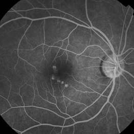 Unilateral PIC Following Recent Influenza Vaccine
Jan 6 2019 by John S. King, MD
FA 5 minute 42-year-old African American female with high myopia and type 2 diabetes, presented to her eye doctor with distortion in the right eye "that looked like seeing through a Coke bottle." She denied any photopsias or other symptoms. She received an influenza vaccine two weeks before onset of metamorphopsia. I saw her about a month after symptoms began. Va cc 20/30 OD J1 (20/15 J1+ OS); a/c and vitreous without cell or flare. Posterior pole OD showed yellowish, rounded small to medium RPE pigment alterations without heme or exudes (OS U/R). FA showed early focal areas of hyperfluorescence that stained in the later frames without CNVM or CME; rare MA inferiorly. The OCT showed some focal PEDs with some possible overlying SRHRM. We discussed options and decided to try a medrol dose pack. A few weeks later she was 20/20 J1, with minimal to no symptoms; OCT shows near total resolution of PEDs.
Photographer: Kay Dalby
Imaging device: Topcon 50
Condition/keywords: punctate inner choroidopathy (PIC), white dot syndrome
-
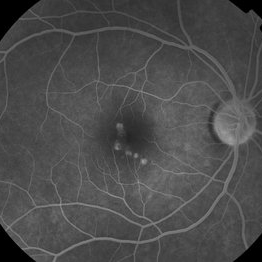 Unilateral PIC Following Recent Influenza Vaccine
Jan 6 2019 by John S. King, MD
FA 2 minute 42-year-old African American female with high myopia and type 2 diabetes, presented to her eye doctor with distortion in the right eye "that looked like seeing through a coke bottle". She denied any photopsias or other symptoms. She received an influenza vaccine two weeks before onset of metamorphopsia. I saw her about a month after symptoms began. Va cc 20/30 OD J1 (20/15 J1+ OS); a/c and vitreous without cell or flare. Posterior pole OD showed yellowish, rounded small to medium RPE pigment alterations without heme or exudes (OS U/R). FA showed early focal areas of hyperfluorescence that stained in the later frames without CNVM or CME; rare MA inferiorly. The OCT showed some focal PEDs with some possible overlying SRHRM. We discussed options and decided to try a medrol dose pack. A few weeks later she was 20/20 J1, with minimal to no symptoms; OCT shows near total resolution of PEDs.
Photographer: Kay Dalby
Imaging device: Topcon 50
Condition/keywords: punctate inner choroidopathy (PIC), white dot syndrome
-
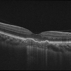 Unilateral PIC Following Recent Influenza Vaccine
Jan 6 2019 by John S. King, MD
Initial OCT. 42-year-old African American female with high myopia and type 2 diabetes, presented to her eye doctor with distortion in the right eye "that looked like seeing through a Coke bottle." She denied any photopsias or other symptoms. She received an influenza vaccine two weeks before onset of metamorphopsia. I saw her about a month after symptoms began. Va cc 20/30 OD J1 (20/15 J1+ OS); a/c and vitreous without cell or flare. Posterior pole OD showed yellowish, rounded small to medium RPE pigment alterations without heme or exudes (OS U/R). FA showed early focal areas of hyperfluorescence that stained in the later frames without CNVM or CME; rare MA inferiorly. The OCT showed some focal PEDs with some possible overlying SRHRM. We discussed options and decided to try a medrol dose pack. A few weeks later she was 20/20 J1, with minimal to no symptoms; OCT shows near total resolution of PEDs.
Photographer: Kay Dalby
Imaging device: Topcon 50
Condition/keywords: punctate inner choroidopathy (PIC), white dot syndrome
-
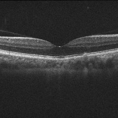 Unilateral PIC Following Recent Influenza Vaccine
Jan 6 2019 by John S. King, MD
OCT few weeks after initial visit. 42-year-old African American female with high myopia and type 2 diabetes, presented to her eye doctor with distortion in the right eye "that looked like seeing through a Coke bottle." She denied any photopsias or other symptoms. She received an influenza vaccine two weeks before onset of metamorphopsia. I saw her about a month after symptoms began. Va cc 20/30 OD J1 (20/15 J1+ OS); a/c and vitreous without cell or flare. Posterior pole OD showed yellowish, rounded small to medium RPE pigment alterations without heme or exudes (OS U/R). FA showed early focal areas of hyperfluorescence that stained in the later frames without CNVM or CME; rare MA inferiorly. The OCT showed some focal PEDs with some possible overlying SRHRM. We discussed options and decided to try a medrol dose pack. A few weeks later she was 20/20 J1, with minimal to no symptoms; OCT shows near total resolution of PEDs.
Photographer: Kay Dalby
Imaging device: Topcon 50
Condition/keywords: punctate inner choroidopathy (PIC), white dot syndrome
A project from the American Society of Retina Specialists