-
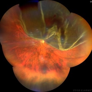
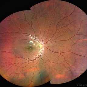
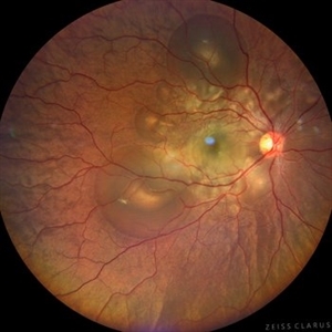
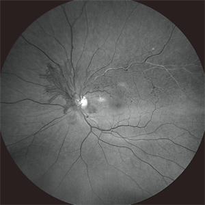
 Post Traumatic Optic Nerve Head Avulsion
Nov 18 2017 by Vishal Agrawal, MD, FRCS,FACS,FASRS
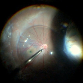
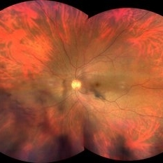
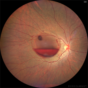
Right eye fundus picture of a 24-year-old male patient who suffered blunt trauma 7 days back with a wooden stick . He presented with NLP vision with a non reacting dilated pupil. Fundus montage picture shows ONH avulsion,CRAO,peripapillary resolving hemorrhages and cicatricial tissue at the edge.
Photographer: Vishal Agrawal, MD, SMS Medical College, Jaipur, India
Imaging device: Zeiss 524
Condition/keywords: avulsion, central retinal artery occlusion (CRAO)
-
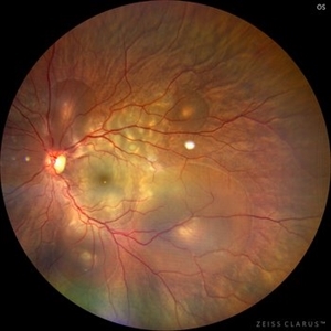
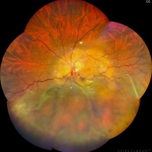
 Familial Exudative Vitreoretinopathy (FEVR)
Apr 14 2018 by Vishal Agrawal, MD, FRCS,FACS,FASRS
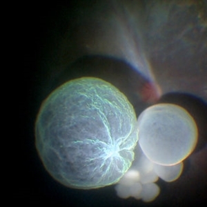
Intraoperative view of a 20-year-old male patient of FEVR operated for a combined tractional and rheghmatogenous RD and macular hole. The right eye image shows temporal fibrovascular band, macular drag, stained inverted flap in the macular hole under the PFCL bubble. Also seen is the nasal indentation mound with its shadow falling over the retina.
Photographer: Vishal Agrawal MD,FRCS
Imaging device: SONY PMW-10 MD HD
Condition/keywords: familial exudative vitreoretinopathy (FEVR)
-
 Intraocular Multiple Cysticercus
Oct 10 2018 by Vishal Agrawal, MD, FRCS,FACS,FASRS
Intraoperative fundus picture of right eye of a 18-year-old boy with complaints of DOV for the past 2 months. There were 12 intravitreal cysts in total with vitritis sclerosis retinal vessels and TRD. To note here, the largest cyst has a flimsy wall and no scolex (possibly ruptured) and the rest of the smaller cysts have a scolex and a taut wall.
Photographer: Vishal Agrawal MD,FRCS
Imaging device: SONY PMW-10 MD HD
Condition/keywords: cysticercosis, scolex
-
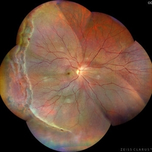
 Retinoblastoma Group C
Dec 29 2019 by Vishal Agrawal, MD, FRCS,FACS,FASRS
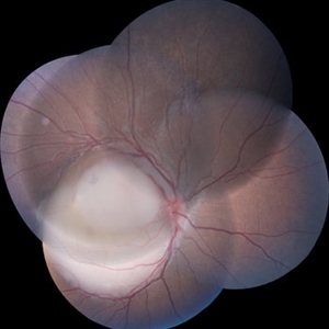
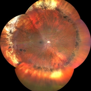
Fundus montage of a 3-year-old boy with mass lesion involving macula and abutting the disc of the right eye.The superior half of the tumor shows preretinal extension.
Photographer: Vishal Agrawal MD
Imaging device: Zeiss
Condition/keywords: retinoblastoma
-
 Choroidal Metastases
Jan 18 2020 by Vishal Agrawal, MD, FRCS,FACS,FASRS
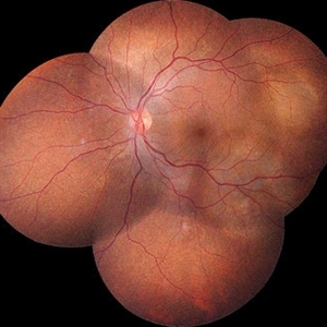
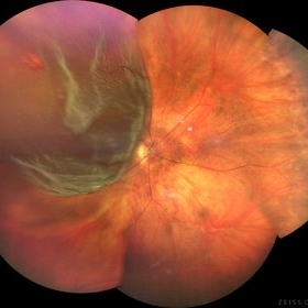
Left eye fundus montage of a 55-year-old female with choroidal metastases with the primary being breast carcinoma. The right eye had exudative retinal detachment.
Photographer: Dr Vishal Agrawal MD,FRCS
Imaging device: Zeiss
Condition/keywords: breast cancer, choroidal metastasis, metastatic lesion
-
 Fundus Fluorescein Angiography of Choroidal Metastases
Jan 18 2020 by Vishal Agrawal, MD, FRCS,FACS,FASRS
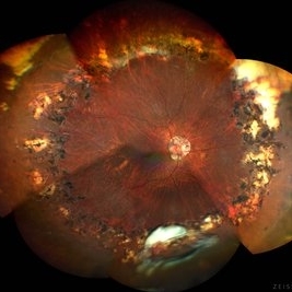
Left eye FFA montage of a 55-year-old female with choroidal metastases with the primary being breast carcinoma. The right eye had exudative retinal detachment . Note the pin point leaks at the border of the 2 lesions.
Photographer: Dr Vishal Agrawal MD,FRCS
Imaging device: Zeiss
Condition/keywords: breast cancer, FA mid phase, metastatic lesion
-
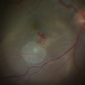 Subretinal Cysticercus Cyst -Intraoperative Picture
Mar 5 2021 by Vishal Agrawal, MD, FRCS,FACS,FASRS
24-year-old male presented with DOV in right for the past 1 month. On examination live cyst noted at macula with inflammatory exudation at the posterior pole.
Photographer: Vishal Agrawal MD,FRCS
Imaging device: SONY PMW-10 MD HD
Condition/keywords: cyst, cysticercosis, macula lesion
-
 Perforating thorn injury
Jun 13 2022 by Vishal Agrawal, MD, FRCS,FACS,FASRS
A 18 year old boy presented with injury by a plant thorn. The thorn entered in stab wound fashion through para plana, falling just short of the retinal surface. The IOFB was removed after PPV and repair of the scleral wound. The image is a still of the intra operative view during vitrectomy.
Photographer: Vishal Agrawal MD
Imaging device: SONY PMW-10 MD HD
Condition/keywords: globe perforation, trauma, vitrectomy
-
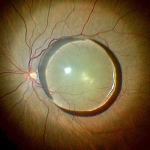 Post traumatic dislocated crystalline lens
Jul 10 2022 by Vishal Agrawal, MD, FRCS,FACS,FASRS
Intra operative picture of a 70 year old female who presented with a history of blunt trauma and vision loss .
Photographer: Vishal Agrawal MD
Imaging device: SONY PMW 10 MD
Condition/keywords: dislocated crystalline lens, retina, trauma
-
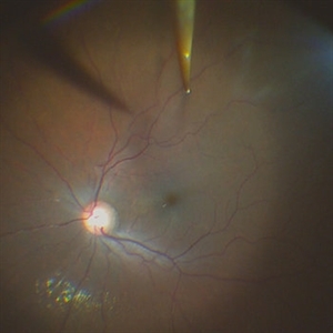
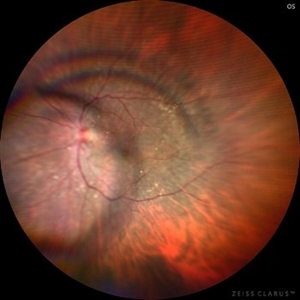
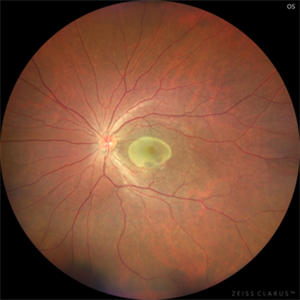
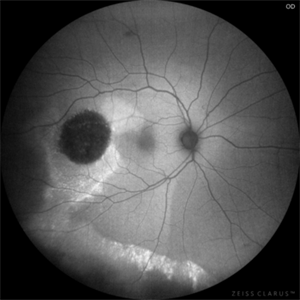
 Optic Disc Melanocytoma
Aug 31 2022 by Vishal Agrawal, MD, FRCS,FACS,FASRS
Optic disc Melanocytoma detected on routine examination in the left eye of a 30 year old male .
Photographer: Vishal Agrawal
Imaging device: Clarus 700
Condition/keywords: Melanocytoma
-
 Retinal Detachment
Sep 9 2022 by Vishal Agrawal, MD, FRCS,FACS,FASRS
24-year-old female patient presented with sudden decrease in vision. On examination there was a left eye subtotal retinal detachment involving macula.
Photographer: Vishal Agrawal MD
Imaging device: Clarus 700
Condition/keywords: bullous retinal detachment, retinal break
-
 Horseshoe Tear with Vitreous Hemorrhage
Oct 5 2022 by Vishal Agrawal, MD, FRCS,FACS,FASRS
Fundus picture of a 54 year old female presenting with acute onset of floaters . On examination nasal HST was noted & lasered.
Photographer: Pankaj
Imaging device: CLARUS 700
Condition/keywords: hemorrhage, tear
-
 Post PPV for retinal detachment
Oct 13 2022 by Vishal Agrawal, MD, FRCS,FACS,FASRS
3 month Post operative picture of right eye .The patient had PPV + gas (C3F8) for total retinal detachment with multiple breaks.
Photographer: Pankaj, Agrawal Hospital,Jaipur
Imaging device: Clarus 700
-
 Giant Retinal Tear
Oct 13 2022 by Vishal Agrawal, MD, FRCS,FACS,FASRS
65 year old female presented with sudden vision loss. On examination superior giant tear with retinal detachment was noted in the right eye.
Photographer: Mahipal,Agrawal Hospital
Imaging device: Clarus 700
Condition/keywords: giant retinal tear, Retinal Detachment
-
 Retinal Detachment with HST.
Oct 22 2022 by Vishal Agrawal, MD, FRCS,FACS,FASRS
Supero-temporal retinal detachment with single horse shoe tear.
Photographer: Pankaj
Imaging device: CLARUS 700
Condition/keywords: Retinal Detachment
-
 Posteriorly dislocated IOL
Oct 22 2022 by Vishal Agrawal, MD, FRCS,FACS,FASRS
67 yr old male , post PPV for retinal detachment ( 5 years ) presented with sudden DOV . On examination posteriorly dislocated 4 loop haptic iol - bag complex was noted .
Photographer: Pankaj
Imaging device: CLARUS 700
Condition/keywords: dropped intraocular lens (IOL)
-
 Stickler syndrome with retinal detachment
Jan 14 2023 by Vishal Agrawal, MD, FRCS,FACS,FASRS
30 year old female with stickler syndrome and subtotal retinal detachment . Fundus pic shows radial para vascular lattices with multiple breaks .
Photographer: Pankaj
Imaging device: CLARUS 700
Condition/keywords: Retinal Detachment, Stickler Syndrome
-
 Superior bullous retinal detachment
Jan 14 2023 by Vishal Agrawal, MD, FRCS,FACS,FASRS
50 year old female with left eye sub total retinal detachment
Photographer: Pankaj
-
 PMMA IOL in mid vitreous cavity - color imaging
Jan 14 2023 by Vishal Agrawal, MD, FRCS,FACS,FASRS
Traumatic dislocation of IOL in vitreous cavity .
Photographer: Pankaj
Imaging device: CLARUS 700
Condition/keywords: posterior dislocation of lens
-
 PMMA IOL in mid vitreous cavity - IR imaging
Jan 14 2023 by Vishal Agrawal, MD, FRCS,FACS,FASRS
Traumatic dislocation of IOL in vitreous cavity .
Photographer: Dr Bhagya Shree
Condition/keywords: dislocated posterior chamber intraocular lens (PCIOL)
-
 Retinal Detachment with multiple breaks
Mar 20 2023 by Vishal Agrawal, MD, FRCS,FACS,FASRS
10 year old female with high myopia presented with right eye retinal detachment.
Photographer: Pankaj
Imaging device: Clarus 700
Condition/keywords: Retinal Detachment
-
 Cone Dystrophy
Aug 30 2023 by Vishal Agrawal, MD, FRCS,FACS,FASRS
12 year old male patient presented with photophobia, decrease in vision and Nystagmus. Bulls eye maculopathy gives an appearance of an eye on the fovea on color fundus photo due to nystagmus.
Photographer: Dr Bhagyashree
Imaging device: Clarus 700
Condition/keywords: bull's eye maculopathy, Cone-Rod Dystrophy
-
 Proliferative Diabetic Retinopathy
Dec 21 2023 by Vishal Agrawal, MD, FRCS,FACS,FASRS
20-year-old male patient having PDR with multiple NVE in all quadrants.
Photographer: Dr Ayushi
Imaging device: Clarus 700
Condition/keywords: Neovascularisation elsewhere (NVE), PDR
-
 Proliferative Diabetic Retinopathy- Red free
Dec 21 2023 by Vishal Agrawal, MD, FRCS,FACS,FASRS
20-year-old male patient having PDR with multiple NVE in all quadrants. Red free image accentuates the NVE as compared to color pic.
Photographer: Dr Ayushi
Imaging device: Clarus 700
Condition/keywords: NVE, PDR, RedFree
-
 Benign Familial Fleck Retina
Dec 21 2023 by Vishal Agrawal, MD, FRCS,FACS,FASRS
10-year male with high myopia on examination revealed diffuse flecks distributed all over fundus in both eyes sparing macula. Inferior lattice with WWOP areas were also noted in right eye.
Photographer: Dr Ayushi
Imaging device: Clarus 700
Condition/keywords: fleck retinopathy, myopia
-
 Benign Familial Fleck Retina
Dec 21 2023 by Vishal Agrawal, MD, FRCS,FACS,FASRS
Green Autoflourescence image of fleck retinopathy.
Photographer: Dr Ayushi
Imaging device: Clarus 700
Condition/keywords: autofluorescence imaging, fleck retinopathy
-
 Central Serous Chorioretinopathy in Pregnancy (OS)
Apr 28 2024 by Vishal Agrawal, MD, FRCS,FACS,FASRS
30-year female with sudden loss of vision came for examination. She was in her first trimester of pregnancy. Examination revealed bilateral bullous NSD with subretinal fibrin s/o CSR.
Photographer: Dr Ayushi
Imaging device: Clarus 700
Condition/keywords: Central Serous Chorioretinopathy (CSR), neurosensory detachment of retina, pregnancy
-
 Central Serous Chorioretinopathy in Pregnancy (OD)
Apr 28 2024 by Vishal Agrawal, MD, FRCS,FACS,FASRS
30-year female with sudden loss of vision came for examination. She was in her first trimester of pregnancy. Examination revealed bilateral bullous NSD with subretinal fibrin s/o CSR.
Photographer: Dr Ayushi
Imaging device: Clarus 700
Condition/keywords: Central Serous Chorioretinopathy (CSR), neurosensory detachment of retina, pregnancy
-
 Exudative Retinal Detachment With Choroidal Metastasis
May 1 2024 by Vishal Agrawal, MD, FRCS,FACS,FASRS
Left eye fundus picture of a 65-year-old female with choroidal metastases and exudative retinal detachment. The patient is under treatment for breast carcinoma.
Photographer: Dr Ayushi
Imaging device: Clarus 700
Condition/keywords: choroidal metastasis, exudative detachment
-
 Dislocated IOL
Jun 27 2024 by Vishal Agrawal, MD, FRCS,FACS,FASRS
Fundus picture of a 65-year-old male patient presenting with posteriorly dislocated IOL & Soemmerring ring in the right eye. PPV + IOL removal + secondary IOL (Yamane technique) was performed.
Photographer: Dr Ayushi
Imaging device: Clarus 700
Condition/keywords: IOL, Soemmering's ring
-
 Retinitis Pigmentosa with PPRPE
Jan 27 2025 by Vishal Agrawal, MD, FRCS,FACS,FASRS
16 year-old male patient presented with DOV, nyctalopia and nystagmus. Fundus revealed pigment clumping, pale disc and preserved para-arteriolar retinal pigment epithelium (PPRPE) in both eyes. Genetic testing revealed CRB1 gene mutation.
Photographer: Dr Ayushi
Imaging device: Clarus 700
Condition/keywords: retinitis pigmentosa
-
 Retinitis Pigmentosa with PPRPE - FAF-G
Jan 27 2025 by Vishal Agrawal, MD, FRCS,FACS,FASRS
16 year-old male patient presented with DOV, nyctalopia and nystagmus. Fundus revealed pigment clumping, pale disc and preserved para-arteriolar retinal pigment epithelium (PPRPE) in both eyes. Genetic testing revealed CRB1 gene mutation.
Photographer: Dr Ayushi Gupta
Imaging device: Clarus 700
Condition/keywords: retinitis pigmentosa
-
 CSR with Fibrin
Jan 28 2025 by Vishal Agrawal, MD, FRCS,FACS,FASRS
A 31-year-old female was referred with a diagnosis of subretinal cysticercosis. BCVA was 20/200 OS. OCT showed a large subfoveal bacillary layer detachment (BALAD) without any scolex. FFA revealed a smoke-stack appearance. A final diagnosis of CSR with Fibrin was made and was managed conservatively. BCVA at final visit was 20/20.
Photographer: Dr Ayushi Gupta
Imaging device: Clarus 700
Condition/keywords: central serous chorioretinopathy (CSCR)
-
 CSR with Fibrin-FFA
Jan 29 2025 by Vishal Agrawal, MD, FRCS,FACS,FASRS
A 31-year-old female was referred with a diagnosis of subretinal cysticercosis. BCVA was 20/200 OS. OCT showed a large subfoveal bacillary layer detachment (BALAD) without any scolex. FFA revealed a smoke-stack appearance. A final diagnosis of CSR with Fibrin was made and was managed conservatively. BCVA at final visit was 20/20.
Photographer: Dr Ayushi Gupta
Imaging device: Clarus 700
Condition/keywords: central serous chorioretinopathy (CSCR)
-
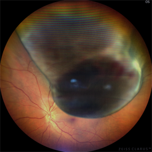
 Sub Hyaloid Hemorrhage
Jan 29 2025 by Vishal Agrawal, MD, FRCS,FACS,FASRS
A 45-year-old male patient presented with decreased vision in Left Eye. On fundus examination, a boat-shaped sub hyaloid hemorrhage was noted. YAG hyaloidotomy was performed and the patient recovered with a vision of 20/20.
Photographer: Dr Ayushi Gupta
Imaging device: Clarus 700
Condition/keywords: YAG HYALOIDOTOMY
-
 FEVR-FFA
Feb 5 2025 by Vishal Agrawal, MD, FRCS,FACS,FASRS
A 22- year male, one eyed patient came for routine examination. Fundus showed temporal straightening of Vessels. FA revealed peripheral avascular area and leakage. 3 siblings had the same findings with no history of prematurity. All the siblings underwent laser treatment.
Photographer: Dr Ayushi Gupta
Imaging device: Clarus 700
Condition/keywords: familial exudative vitreoretinopathy (FEVR)
-
 FEVR
FEVR
Feb 6 2025 by Vishal Agrawal, MD, FRCS,FACS,FASRS
A 22- year male, one eyed patient came for routine examination. Fundus showed temporal straightening of Vessels. FA revealed peripheral avascular area and leakage. 3 siblings had the same findings with no history of prematurity. All the siblings underwent laser treatment.
Photographer: Dr Ayushi Gupta
Imaging device: Clarus 700
Condition/keywords: familial exudative vitreoretinopathy (FEVR)
-
 Subhyaloid Hemorrhage
Mar 1 2025 by Vishal Agrawal, MD, FRCS,FACS,FASRS
A 37-year-old male presented with sudden diminution of vision in the right eye. On fundus examination boat shaped sub hyaloid hemorrhage was noted and a YAG hyaloidotomy was performed.
Photographer: Dr Ayushi Gupta
Imaging device: Clarus 700
Condition/keywords: YAG HYALOIDOTOMY
-
 FAF-G Circumscribed Choroidal Hemangioma
Mar 1 2025 by Vishal Agrawal, MD, FRCS,FACS,FASRS
A 37-year-old male presented with decreased vision in the right eye. This is the fundus autofluorescence (FAF-G) of the right eye showing hypo auto fluorescent lesion with surrounding hyper auto fluorescence extending inferiorly corresponding to the fluid tract.
Photographer: Dr Ayushi Gupta
Imaging device: Clarus 700
Condition/keywords: Circumscribed Choroidal Hemangioma, fundus autofluorescence (FAF)
-
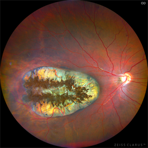
 Uveal Melanoma
Apr 26 2025 by Vishal Agrawal, MD, FRCS,FACS,FASRS
A 32 year-old male presented with complaints of perceiving a shadow in OS for 15-20 days. His BCVA was 20/20 OU. On Fundus examination, a large, elevated, well-defined, pigmented choroidal mass with few hemorrhages over the lesion was seen and a provisional diagnosis of uveal melanoma was made. urgent oncological consultation was recommended for further treatment.
Photographer: Dr Ayushi Gupta
Imaging device: Clarus 700
Condition/keywords: melanoma
-
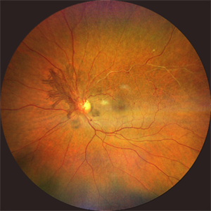 Neovascularization of the Disc (NVD)
Apr 28 2025 by Vishal Agrawal, MD, FRCS,FACS,FASRS
Fundus image showing prominent neovascularization of the disc (NVD)- visible as fine, frond-like vascular proliferation extending from the disc surface.
Photographer: Dr Ayushi Gupta
Imaging device: Clarus 700
Condition/keywords: branch retinal vein occlusion (BRVO), NVD
-
 Neovascularization of the Disc (NVD) - Red Free
Apr 28 2025 by Vishal Agrawal, MD, FRCS,FACS,FASRS
The red-free image enhances the visualization of the NVD, showing the fine neovascular fronds sprouting from the optic disc. Collateral vessels and vascular anastomosis are better appreciated.
Photographer: Dr Ayushi Gupta
Imaging device: Clarus 700
Condition/keywords: branch retinal vein occlusion (BRVO), collaterals
-
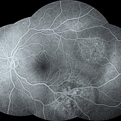
 Takayasu Retinopathy
Apr 30 2025 by Vishal Agrawal, MD, FRCS,FACS,FASRS
Fundus fluorescein angiography image of a young girl with diagnosed Takayasu arteritis who presented with complains of diminished vision in both eyes. FFA shows complete absence of venous filling with segmented blood column secondary to CRAO with peripheral avascular area.
Photographer: Dr Ayushi Gupta
Imaging device: Clarus 700
Condition/keywords: calcified drusen, CRAO, takayasu arteritis
-
 Takayasu Retinopathy
Apr 30 2025 by Vishal Agrawal, MD, FRCS,FACS,FASRS
Fundus fluorescein angiography image of a young girl with diagnosed Takayasu arteritis who presented with complains of diminished vision in both eyes. FFA shows complete absence of venous filling with segmented blood column secondary to CRAO with peripheral avascular area.
Photographer: Dr Ayushi Gupta
Imaging device: Clarus 700
Condition/keywords: CRAO, Takayasus disease
-
 Presumed Congenital Toxoplasmosis
Aug 16 2025 by Vishal Agrawal, MD, FRCS,FACS,FASRS
Fundus picture of 7 a year-old boy with esotropia. OCT showed complete atrophy & disorganization of the overlying RPE and neurosensory retina.
Photographer: Dr Ayushi Gupta
Imaging device: Clarus 700
Condition/keywords: coloboma of macula, toxoplasmosis
-
 Presumed Congenital Toxoplasmosis Macular Coloboma
Aug 16 2025 by Vishal Agrawal, MD, FRCS,FACS,FASRS
7-year-old boy presented with esotropia in OD with light perception positive. Fundus reveals a large macular coloboma occupying nearly the entire macula. OCT scan shows complete atrophy and disorganization of the overlying RPE and neurosensory retina. A much smaller lesion was observed in OS with BCVA 20/40.
Photographer: Dr Ayushi Gupta
Imaging device: Clarus 700
Condition/keywords: Coloboma, congenital toxoplasmosis
-
 Choroidal Rupture
Dec 4 2025 by Vishal Agrawal, MD, FRCS,FACS,FASRS
Color fundus pic of Left Eye showing curvilinear, crescent-shaped choroidal rupture, extending across the macular region, post blunt trauma. The rupture appears bright, yellow-white streak with well-delineated margins, consistent with exposed underlying sclera due to disruption of the choriocapillaris and Bruch's membrane.
Photographer: Dr Ayushi Gupta, Agrawal Hospital, Jaipur
Imaging device: Clarus 700
Condition/keywords: blunt trauma, choroidal rupture
-
 Myelinated Nerve Fiber
Dec 5 2025 by Vishal Agrawal, MD, FRCS,FACS,FASRS
Fundus picture of left eye shows dense area of myelinated retinal nerve fibers involving the peripapillary region and extending along the superior and inferior temporal arcades.
Photographer: Dr Ayushi Gupta, Agrawal Hospital, Jaipur
Imaging device: Clarus 700
Condition/keywords: amblyopia, myelinated nerve fiber layer
A project from the American Society of Retina Specialists