-
 CNV due to AMPPE
Oct 16 2012 by Ratimir Lazic, MD, PhD
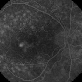
FAG of 58-year-old male. In late venous phase hyperflorescence of white dots (caused by window defect) can be seen. Intensive leakage of dye in juxtafoveolar region.
Photographer: Marko Lukic, MD
Imaging device: Zeis Visucam Lite 2
Condition/keywords: acute posterior multifocal placoid pigment epitheliopathy (APMPPE), choroidal neovascularization (CNV)
-
 AMPPE
AMPPE
Oct 16 2012 by Ratimir Lazic, MD, PhD
Color fundus photography of a 58 -year-old male. Yellow- white placoid dots can be seen in macular region. BCVA of that eye is 0.9.
Photographer: Marko Lukic, MD
Imaging device: Zeis Visucam Lite 2
Condition/keywords: acute posterior multifocal placoid pigment epitheliopathy (APMPPE)
-
 CNV due to AMPPE
Oct 16 2012 by Ratimir Lazic, MD, PhD
Color fundus photography of a 58-year-old male. White dots with juxtafoveolar subretinal fluid can be seen. BCVA of that eye is 0.35.
Photographer: Marko Lukic, MD
Imaging device: Zeis Visucam Lite 2
Condition/keywords: acute posterior multifocal placoid pigment epitheliopathy (APMPPE), choroidal neovascularization (CNV)
-
 CNV due to AMPPE
Oct 16 2012 by Ratimir Lazic, MD, PhD
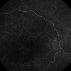
FAG of 58-year-old male. In early venous phase hyperflorescence of white dots (caused by window defect) can be seen. Leakage of dye in juxtafoveolar region.
Photographer: Marko Lukic, MD
Imaging device: Zeis Visucam Lite 2
Condition/keywords: acute posterior multifocal placoid pigment epitheliopathy (APMPPE), choroidal neovascularization (CNV)
-
 CNV due to AMPPE
Oct 16 2012 by Ratimir Lazic, MD, PhD
OCT image of 58-year- old male. Total resolution of fluid one and a half month after treatment can be seen. The patient was treated with intravitreal bevacizumab.
Photographer: Marko Lukic, MD
Imaging device: OCT Copernicus
Condition/keywords: acute posterior multifocal placoid pigment epitheliopathy (APMPPE), anti-VEGF, choroidal neovascularization (CNV)
A project from the American Society of Retina Specialists