-
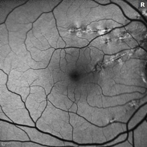 Idiopathic Choroidal Folds
Aug 22 2017 by Carolyn Daley
This is an autofluorescence image of a 77-year-old male with idiopathic choroidal folds in his right eye.
Photographer: Carolyn Daley
Imaging device: Heidelberg Spectralis
Condition/keywords: autofluorescence imaging, choroidal folds
-
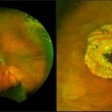
 UWF of Retinal Detachment Corrected with Scleral Buckle
Aug 29 2017 by Carolyn Daley
An ultra wide field fundus photograph of a 57-year-old male who has a past history of retinal detachment corrected with scleral buckle and three treated retinal tears.
Photographer: Carolyn Daley
Imaging device: Optos Imaging
Condition/keywords: cryo-retinal tear, cryotherapy, Optos, retinal tear, scleral buckle, ultra-wide field imaging
-
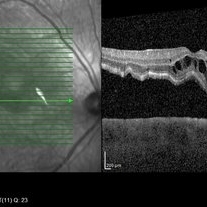
 OCT Image of Epiretinal Membrane
Aug 29 2017 by Carolyn Daley
OCT photograph of a 64-year-old women with an epiretinal membrane in the right eye. Patient has not noticed any decline in vision so surgery was not recommended at this time.
Photographer: Carolyn Daley
Imaging device: Heidelberg Spectralis
Condition/keywords: epiretinal membrane (ERM), optical coherence tomography (OCT)
-
 Macula Off Retinal Detachment
Jan 2 2018 by Carolyn Daley
55-year-old with macula off retinal detachment post cataract surgery.
Photographer: Carolyn Daley, Retina Specialists of Michigan
Imaging device: Heidelberg Spectralis
Condition/keywords: Heidelburg Spectralis, optical coherence tomography (OCT)
-
 Operculated Hole and CHRPE
Jan 16 2018 by Carolyn Daley
58-year-old woman with an operculated hole and CHRPE in the right eye. Patient is asymptomatic so no treatment was recommended at this time.
Photographer: Carolyn Daley
Imaging device: Optos ultra wide field image
Condition/keywords: congenital hypertrophy of the retinal pigment epithelium (CHRPE), operculated retinal hole, Optos, ultra-wide field imaging
-
 Lattice Degeneration
Apr 27 2018 by Carolyn Daley
Fundus photograph of a 15-year-old woman with lattice degeneration and atrophic holes. Patient will have laser treatment at her next appointment.
Photographer: Carolyn Daley, Retina Specialists of Michigan
Imaging device: OPTOS Ultra-Wide Field Camera
Condition/keywords: atrophic retinal hole, lattice degeneration, Optos
-
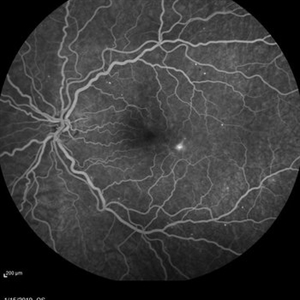 Mild Nonproliferative Diabetic Retinopathy
Jan 16 2019 by Carolyn Daley
Fluorescence angiogram 50 degree imaging of a 38-year-old woman with mild nonproliferative diabetic retinopathy in the left eye. Patient also presented with a paracentral scotoma which etiologies could include vascular occlusion vs JFRT.
Photographer: Carolyn Daley, Retina Specialists of Michigan
Imaging device: Heidelberg Spectralis
Condition/keywords: diabetes, Heidelburg Spectralis, juxtafoveal telangiectasis, nonproliferative diabetic retinopathy, retinopathy, vascular occlusions
-
 Choroidal Melanoma
May 21 2019 by Carolyn Daley
Optos ultra-wide field imaging of a 48-year-old man with a choroidal melanoma in the left eye.
Photographer: Carolyn Daley, COA, Retina Specialists of Michigan
Imaging device: Optos ultra-wide field imaging
Condition/keywords: autofluorescence imaging, Optos, ultra-wide field imaging
A project from the American Society of Retina Specialists