-
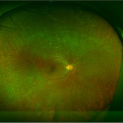
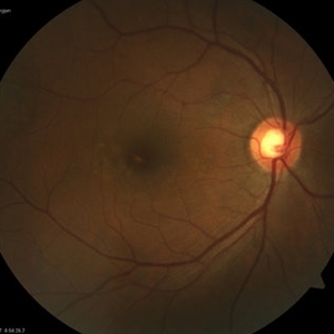 Retinal Pigment Epithelitis
Jul 31 2017 by Eitae Kim, MD
Fundus photograph which shows yellow-whitish dot at subfovea.
Photographer: Eitae Kim, BOIM retinal center, Pureun eye hospital
Imaging device: Visucam, Zeiss
Condition/keywords: fundus photograph, retinal pigment epithelium
-
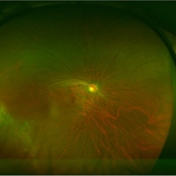 Wagner Syndrome
Aug 1 2017 by Eitae Kim, MD
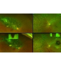
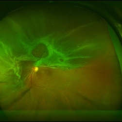
Ultra wide field fundus photograph of 19-year-old male with Wagner syndrome.
Photographer: Eitae Kim, BOIM retinal center, Pureun eye hospital
Condition/keywords: ultra-wide field imaging, Wagner disease
-
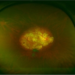
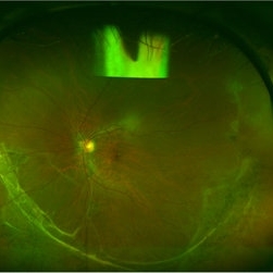 Wagner Syndrome
Aug 1 2017 by Eitae Kim, MD
Ulltra wide field fundus photograph of 19-year-old male with Wagner syndrome which shows peripheral subretinal fibrosis and pigmentary degeneration.
Photographer: Eitae Kim, BOIM retinal center, Pureun eye hospital
Condition/keywords: subretinal fibrosis, ultra-wide field imaging, Wagner disease
-
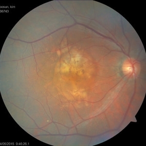 Large PED in AMD, First Visit
Aug 1 2017 by Eitae Kim, MD
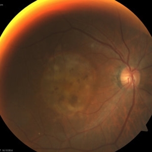
Initial fundus photograph of 78-year-old male with advanced AMD.
Photographer: Eitae Kim, BOIM retinal center, Pureun eye hospital
Imaging device: Visucam, Zeiss
Condition/keywords: age-related macular degeneration (AMD), fundus photograph
-
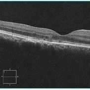
 Large PED in AMD, First Visit
Aug 1 2017 by Eitae Kim, MD
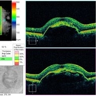
Initial OCT of 78-year-old male with advanced AMD.
Photographer: Eitae Kim, BOIM retinal center, Pureun eye hospital
Imaging device: Cirrus, Zeiss
Condition/keywords: optical coherence tomography (OCT)
-
 Large PED in AMD, 2 Years After
Aug 1 2017 by Eitae Kim, MD
Fundus photograph of 78-year-old male with advanced AMD after 6 injections of bevacizumab.
Photographer: Eitae Kim, BOIM retinal center, Pureun eye hospital
Imaging device: Visucam, Zeiss
Condition/keywords: fundus photograph
-
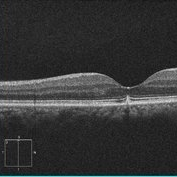
 Large PED in AMD, 2 Years After
Aug 1 2017 by Eitae Kim, MD
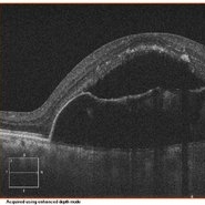
OCT of 78-year-old male with advanced AMD after 6 injections of bevacizumab.
Photographer: Eitae Kim, BOIM retinal center, Pureun eye hospital
Imaging device: Cirrus, Zeiss
Condition/keywords: optical coherence tomography (OCT)
-
 Large PED in AMD, 2 Years After
Aug 1 2017 by Eitae Kim, MD
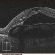
OCT of 78-year-old male with advanced AMD after 6 injections of bevacizumab.
Photographer: Eitae Kim, BOIM retinal center, Pureun eye hospital
Imaging device: Cirrus, Zeiss
Condition/keywords: optical coherence tomography (OCT)
-
 Severe Fibrovascular Proliferative Mass With Tractional RD in PDR
Aug 1 2017 by Eitae Kim, MD
Severe fibrovascular proliferation with tractional retinal detachment is seen on UWF fundus photograph.
Photographer: Eitae Kim, BOIM retinal center, Pureun eye hospital
Condition/keywords: fibrous proliferation, tractional retinal detachment
-
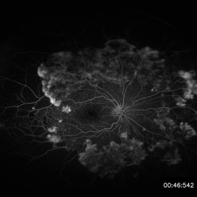
 Severe Fibrovascular Proliferative Mass With Tractional RD in PDR
Aug 1 2017 by Eitae Kim, MD
Severe fibrovascular proliferation with tractional retinal detachment is seen on UWF FAG.
Photographer: Eitae Kim, BOIM retinal center, Pureun eye hospital
Condition/keywords: fibrous proliferation, tractional retinal detachment
-
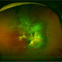
 Sea Fan Like Severe Fibrovascular Proliferation is Seen on Ultra Wide Field Fundus Photograph
Aug 1 2017 by Eitae Kim, MD
Sea fan like severe fibrovascular proliferation on UWF fundus photograph of 55-year-old male with PDR before treatment.
Photographer: Eitae Kim, BOIM retinal center, Pureun eye hospital
Condition/keywords: fibrovascular proliferation, proliferative diabetic retinopathy (PDR), sea fan
-
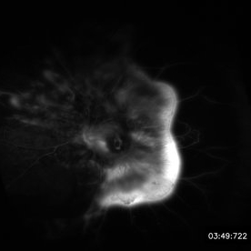
 Sea Fan Like Severe Fibrovascular Proliferation is Seen on Ultra Wide Field FAG
Aug 1 2017 by Eitae Kim, MD
UWF FAG image of 55-year-old male with PDR before treatment.
Photographer: Eitae Kim, BOIM retinal center, Pureun eye hospital
Condition/keywords: fibrovascular proliferation, proliferative diabetic retinopathy (PDR), sea fan
-
 Regressed PDR After PPV
Aug 1 2017 by Eitae Kim, MD
Severe tractional retinal detachment with fibrovascular proliferation regressed after PPV and silicone oil injection.
Photographer: Eitae Kim, BOIM retinal center, Pureun eye hospital
Condition/keywords: ultra-wide field imaging
-
 Regressed PDR After PRP and Bevacizumab Injection
Aug 1 2017 by Eitae Kim, MD
Severe new vessels in PDR dramatically regressed after PRP and bevacizumab injection.
Photographer: Eitae Kim, BOIM retinal center, Pureun eye hospital
Condition/keywords: fundus photograph
-
 Retinal Pigment Epithelitis
Aug 1 2017 by Eitae Kim, MD
OCT image of 51-year-old male with recurrent retinal pigment epithelitis.
Photographer: Eitae Kim, BOIM retinal center, Pureun eye hospital
Imaging device: Cirrus, Zeiss
Condition/keywords: optical coherence tomography (OCT), retinal pigment epithelium
-
 Retinal Pigment Epithelitis
Aug 1 2017 by Eitae Kim, MD
OCT image which shows peaked RPE layer at subfoveal region in retinal pigment epithelitis.
Photographer: Eitae Kim, BOIM retinal center, Pureun eye hospital
Imaging device: Cirrus, Zeiss
Condition/keywords: optical coherence tomography (OCT), retinal pigment epithelium
-
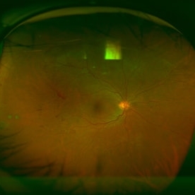
 Posterior Staphyloma
Aug 3 2017 by Eitae Kim, MD
UWF fundus photograph of 35-year-old male with unilateral posterior staphyloma.
Photographer: Eitae Kim, BOIM retina center, Pureun eye hospital
Condition/keywords: ultra-wide field imaging
-
 Bilateral Macroaneurysm
Aug 4 2017 by Eitae Kim, MD
UWF fundus photograph of 81-year-old woman with diabetic retinopathy. Recently the blood pressure was abnormally high and uncontrolled. The above is 6-month- ago fundus photograph and below is recent photograph.
Photographer: Eitae Kim, BOIM retina center, Pureun eye hospital
Condition/keywords: ultra-wide field imaging
-
 Combined RD in PDR patient
Sep 8 2017 by Eitae Kim, MD
52-year-old woman with diabetes visited visual disturbance. Fundus exam shows superior tractional-rhegmatogenous retinal detachment with proliferative vitreoretinopathy. I performed PPV, membranectomy, endolaser and silicone oil injection.
Photographer: Eitae Kim, BOIM retina center, Pureun eye hospital
Condition/keywords: ultra-wide field imaging
A project from the American Society of Retina Specialists