-
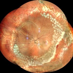
 Pigmentary Paravenous Retinochoroidal Atrophy (Left Eye)
Jun 30 2017 by Navneet Mehrotra, DNB
Fundus photograph of the left eye of a 44-year-old female with bony corpuscle pigmentation along the retinal vessels.
Photographer: Rakesh Juneja, Retina Foundation, Ahmedabad
Condition/keywords: atrophy, pigment, retinochoroidopathy
-
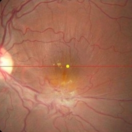
 Pigmentary Paravenous Retinochoroidal Atrophy (Right Eye)
Jun 30 2017 by Navneet Mehrotra, DNB
Fundus photograph of the right eye of a 44-year-old female with bony corpuscle pigmentation along the retinal vessels.
Photographer: Rakesh Juneja, Retina Foundation, Ahmedabad
Condition/keywords: atrophy, retinochoroidopathy
-
 Leber's Miliary Aneurysm With Epiretinal Membrane
Sep 14 2017 by Navneet Mehrotra, DNB
Fundus photo of a 47-year-old male with miliary aneurysms with epiretrinal membrane.
Photographer: Rakesh Juneja, retina foundation Ahmedabad
Condition/keywords: Leber's miliary aneurysm
-
 FA ICG AZOOR
Oct 14 2017 by Navneet Mehrotra, DNB
fundus autofluorescence OD showing peripapillary hypoautofluorescence surrounded by an area of hyperautofluorescence with well demarcated margins suggestive of AZOOR.
Photographer: Ashish jain, Retina Foundation, Ahmedabad
Imaging device: Heidelberg spectralis
Condition/keywords: acute zonal occult outer retinopathy (AZOOR)
-
 FA ICG AZOOR
Oct 14 2017 by Navneet Mehrotra, DNB
Fundus autofluorescence OS showing peripapillary hypoautofluorescence surrounded by an area of hyperautofluorescence with well demarcated margins suggestive of AZOOR.
Photographer: Ashish jain, Retina Foundation, Ahmedabad
Imaging device: Heidelberg spectralis
Condition/keywords: acute zonal occult outer retinopathy (AZOOR)
-
 Progressive Outer Retinal Necrosis
Nov 19 2017 by Navneet Mehrotra, DNB
42-year-old HIV positive male with reduced vision in right eye for two weeks.
Photographer: Mehul Prajapati
Imaging device: Topcon
Condition/keywords: HIV, progressive outer retinal necrosis (PORN)
-
 Bullous RD With Dislocated Lens
Apr 3 2018 by Navneet Mehrotra, DNB
Dislocated clear lens and associated retinal detachment in a young patient with Marfan's syndrome.
Photographer: Navneet Mehrotra
Imaging device: Sony 3 chip camera
Condition/keywords: dislocated crystalline lens, Marfan's syndrome
-
 Post Traumatic ERM With Large Retinal Tear
Apr 9 2018 by Navneet Mehrotra, DNB
A 22-year-old male presented with epiretinal membrane with large retinal tear and pigmentary changes, two months following blunt trauma
Photographer: Mehul Choudhary
Condition/keywords: epiretinal membrane (ERM), pigment changes, retinal tear, trauma
-
 Spontaneously Reattached Retina
Apr 19 2018 by Navneet Mehrotra, DNB
Fundus photograph of a 34-years-old female with spontaneously reattached retina.
Photographer: Mehul Choudhary
Imaging device: Topcon
Condition/keywords: spontaneous retinal reattachment
-
 Small subILM Hemorrhage
Oct 26 2019 by Navneet Mehrotra, DNB
44-year-old hypertensive male with sudden decrease in vision showing small sub ILM hemorrhage at macula.
Photographer: Navneet Mehrotra
Imaging device: NidekRS330
Condition/keywords: hypertension, subILM hemorrhage
-
 Lymphoma Lesion at the Posterior Pole
Dec 27 2019 by Navneet Mehrotra, DNB
Fundus photograph of a 54-year-old female with posterior pole leopard skin lesion temporal to fovea. She underwent treatment for primary CNS lymphoma.
Photographer: Mitanshi
Imaging device: NIDEK MIRANTE
Condition/keywords: lymphoma, posterior pole lesion
-
 Large Posterior Tear Following Trauma
Dec 27 2019 by Navneet Mehrotra, DNB
A 18-year-old male presented with diminution of vision following blunt trauma to the eye. large tear was noted at the posterior pole.
Photographer: Navneet Mehrotra
Imaging device: Topcon
Condition/keywords: posterior tear
-
 Neuroretinitis
Feb 19 2020 by Navneet Mehrotra, DNB
Fundus photograph of a 31-year-old male with blurring of vision for 10 days. History of fever 10 days back. No history of contact with pets.
Photographer: Mitanshi, Retina Foundation, Ahmedabad
Imaging device: NIDEK MIRANTE
Condition/keywords: neuroretinitis
-
 Doyne Honeycomb Retinal Dystrophy
Sep 29 2020 by Navneet Mehrotra, DNB
Left eye fundus photograph of a 36-year-old female with decreased vision both eyes for six months. Father also had a similar retinal disorder.
Photographer: Dr Navneet Mehrotra
Imaging device: TRC- NW8F
Condition/keywords: Doyne's Honeycomb, drusen, Malattia Leventinese
-
 Doyne Honeycomb Retinal Dystrophy
Sep 29 2020 by Navneet Mehrotra, DNB
Right eye fundus photograph of a 36-year-old female with decreased vision both eyes for six months. Father also had a similar retinal disorder.
Photographer: Dr Navneet Mehrotra, Retina Care, Ahmedabad
Imaging device: TRC- NW8F
Condition/keywords: Doyne's Honeycomb, familial drusen, Malattia Leventinese
-
 Sub ILM Bleed in a Case of Retinal Arterial Macroaneurysm
Mar 11 2021 by Navneet Mehrotra, DNB
Fundus photograph of a 42-year-old female, who presented with sudden diminution of vision since morning.
Photographer: Navneet Mehrotra
Imaging device: Nidek RS 330
Condition/keywords: retinal arterial macroaneurysm
-
 Attached Retina in a Silicon Oil Filled Buckled Eye with Retinectomy
Apr 17 2021 by Navneet Mehrotra, DNB
Fundus photograph of a 12-year-old boy operated for re retinal detachment with PVR showing attached retina with fresh and old laser marks, silicon oil filled and relaxing retinectomy.
Photographer: Dr Nivesh Gupta, Retina Foundation
Imaging device: Nidek mirante
Condition/keywords: proliferative vitreoretinopathy (PVR), retinectomy, scleral buckle
-
 Combined hamartoma of retina and retinal pigment epithelium
Aug 8 2023 by Navneet Mehrotra, DNB
A 20 year old female presented with decreased vision and metamorphopsia noticed in her left eye for one year. Other eye was normal. BCVA was 6/12 in her left eye.
Photographer: Dharti, Retina Care , Ahmedabad
Condition/keywords: Combined pigment epithelial and retinal hamartoma
-
 Melanocytoma of the optic disc
Oct 10 2023 by Navneet Mehrotra, DNB
melanocytoma of the optic nerve head in a 48 year old female diagnosed on routine examination
Photographer: Dharti, Retina Care , Ahmedabad
Imaging device: Nidek RS 330
Condition/keywords: benign pigmented lesion, melanocytoma
-
 Left Eye Acute Retinal Pigment Epithelitis
Jul 25 2024 by Navneet Mehrotra, DNB
Fundus photograph of a 17 year old boy with sudden decrease in vision in his left eye. Retinal pigment epithelial changes were noticed at the macula.
Photographer: Navneet Mehrotra
Imaging device: Nidek
Condition/keywords: retinal pigment epithelium (RPE) changes
A project from the American Society of Retina Specialists