-
 X-Linked Juvenile Retinoschisis
Dec 16 2016 by Young Hee Yoon, MD, PhD
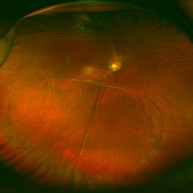
Ultrawide field fundus photograph of a 27-year-old male with x-linked juvenile retinoschisis shows peripheral retinoshisis in his right eye
Photographer: Yoo Jin Jang, University of Ulsan, Asan Medical Center, Seoul, Korea
Imaging device: Optomap
Condition/keywords: juvenile retinoschisis
-
 X-Linked Juvenile Retinoschisis
Dec 16 2016 by Young Hee Yoon, MD, PhD
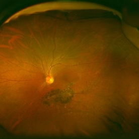
Ultrawide field fundus photograph of a 27-year-old male with x-linked juvenile retinoschisis shows peripheral retinoshisis in his left eye
Photographer: Yoo Jin Jang, University of Ulsan, Asan Medical Center, Seoul, Korea
Imaging device: Optomap
Condition/keywords: juvenile retinoschisis
-
 X-Linked Juvenile Retinoschisis
Dec 17 2016 by Young Hee Yoon, MD, PhD
Optical coherence tomograph of a 27-year-old male with x-linked juvenile retinoschisis shows foveal retinoshisis in his left eye and inferior inner retinal schisis in both eyes.
Photographer: Jin Soo Yeom, University of Ulsan, Asan Medical Center, Seoul, Korea
Imaging device: Spectralis
Condition/keywords: foveal schisis, inferotemporal retinoschisis, juvenile retinoschisis
-
 X-Linked Juvenile Retinoschisis
Dec 17 2016 by Young Hee Yoon, MD, PhD
Fundus photograph of a 27-year-old male with x-linked juvenile retinoschisis shows foveal retinoshisis with spokewheel pattern in his left eye.
Photographer: Ji Soo Kim, University of Ulsan, Asan Medical Center, Seoul, Korea
Condition/keywords: foveal schisis, spokewheel pattern
A project from the American Society of Retina Specialists