-
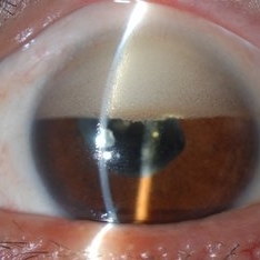 Inverse Hypopyon
Mar 4 2018 by Yoshihiro Yonekawa, MD, FASRS
Slit lamp photograph of a 40-year-old man with previous retinal detachment surgery with silicone oil tamponade, presenting with an inverse hypopyon from emulsified silicone oil.
Photographer: Steven A Bennett, COA, CRA
Imaging device: Nikon D200 / Topcon Slit lamp
Condition/keywords: hypopyon, silicone oil
-
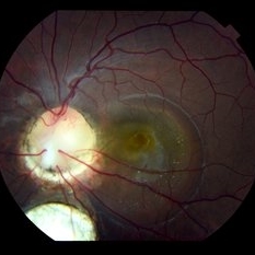 Colobomatous Optic Disc Maculopathy
Feb 13 2020 by Yoshihiro Yonekawa, MD, FASRS
Beautifully focused fundus photograph of a teenage girl with submacular fluid from a colobomatous optic disc.
Photographer: Netanya Lerner, COA, Wills Eye Hospital/Mid Atlantic Retina
Imaging device: Topcon
Condition/keywords: chorioretinal coloboma, coloboma of optic disc, congenital optic nerve pit, subretinal fluid
-
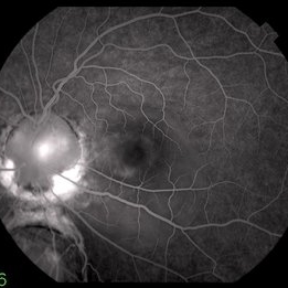 Colobomatous Optic Disc Maculopathy
Feb 13 2020 by Yoshihiro Yonekawa, MD, FASRS
Fluorescein angiography, late frame, of a teenage girl with submacular fluid from a colobomatous optic disc. The camera is focused is on the elevated macula, and the disc is subtly defocused.
Photographer: Netanya Lerner, COA, Wills Eye Hospital/Mid Atlantic Retina
Imaging device: Topcon
Condition/keywords: chorioretinal coloboma, coloboma of optic disc, congenital optic nerve pit, subretinal fluid
-
 Colobomatous Optic Disc Maculopathy
Feb 13 2020 by Yoshihiro Yonekawa, MD, FASRS
EDI-OCT of a teenage girl with submacular fluid from a colobomatous optic disc. Note the subtle tracking of the subretinal fluid into the disc.
Photographer: Netanya Lerner, COA, Wills Eye Hospital/Mid Atlantic Retina
Imaging device: Topcon
Condition/keywords: chorioretinal coloboma, coloboma of optic disc, congenital optic nerve pit, subretinal fluid
-
 Torpedo Maculopathy
Jul 29 2020 by Yoshihiro Yonekawa, MD, FASRS
Fundus photograph of a 10-year-old boy with an incidentally identified torpedo maculopathy.
Photographer: Suely Bascope
Imaging device: Topcon
Condition/keywords: macula lesion, pediatric retina, torpedo maculopathy
-
 Asteroid Hyalosis
Sep 15 2020 by Yoshihiro Yonekawa, MD, FASRS
Fundus photograph of a 70-year-old man with glistening asteroid hyalosis.
Photographer: Alexa Bednar, Mid Atlantic Retina
Imaging device: Topcon
Condition/keywords: asteroid hyalosis
-
 Morning Glory Disc Anomaly
Nov 11 2020 by Yoshihiro Yonekawa, MD, FASRS
Color fundus photograph of a young boy with morning glory disc anomaly. Notice the concavity surrounding the enlarged disc, radial vasculature, and nasally dragged macula. MRI was negative for moyamoya disease, a known association.
Photographer: Alicia Thresher, Mid Atlantic Retina
Imaging device: Topcon
Condition/keywords: disc coloboma, Morning Glory Syndrome, pediatric retina
-
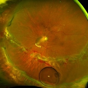 Methotrexate Bubble in Silicone Oil Filled Eye: Proliferative Vitreoretinopathy Prevention
Jan 22 2022 by Yoshihiro Yonekawa, MD, FASRS
A middle-aged man underwent complex retinal detachment repair with vitrectomy, membrane peeling, relaxing retinectomy, and silicone oil tamponade. This is a wide-field image immediately after methotrexate injection during a postoperative clinic visit, for the prevention of proliferative vitreoretinopathy. The methotrexate bubble is seen inferiorly.
Photographer: Christina Rowland
Imaging device: Optos California
Condition/keywords: methotrexate, proliferative vitreoretinopathy (PVR), retinectomy, vitrectomy
A project from the American Society of Retina Specialists