-
 Retinal folds following retinal reattachment surgery
Nov 22 2015 by Mallika Goyal, MD
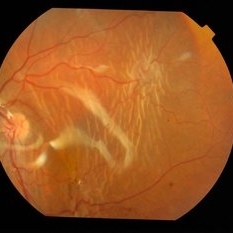
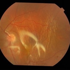
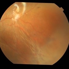
Multiple retinal folds 4 weeks following vitreous surgery (perfluorodecalin assisted) for retinal detachment with giant retinal tear.
Photographer: Mallika Goyal, MD, Apollo Health City, Jubilee Hills, Hyderabad, India
Condition/keywords: retinal fold
-
 Retinal Folds Following Retinal Reattachment Surgery
Nov 22 2015 by Mallika Goyal, MD
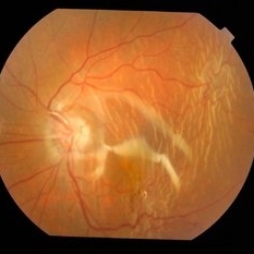
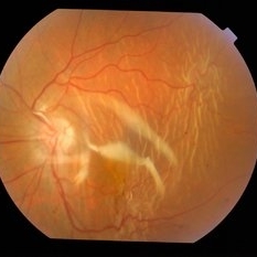
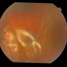
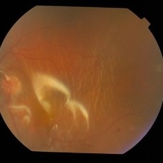
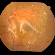
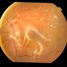
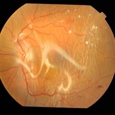
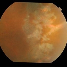
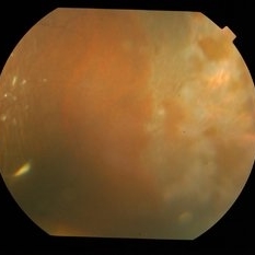
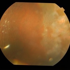
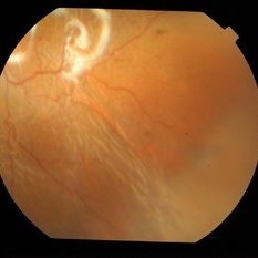
Multiple retinal folds seen in the left part of the fundus photograph 4 weeks following vitreous surgery (perfluorodecalin assisted) for retinal detachment with giant retinal tear. Right part of the figure shows laser scarring for the giant tear.
Photographer: Mallika Goyal, MD, Apollo Health City, Jubilee Hills, Hyderabad, India
Condition/keywords: retinal fold
-
 Retinal Folds Following Retinal Reattachment Surgery
Nov 22 2015 by Mallika Goyal, MD
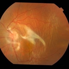
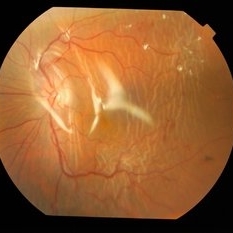
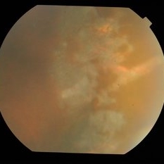
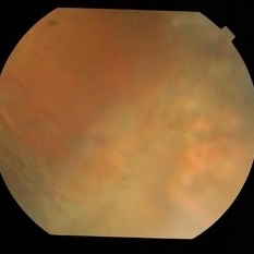
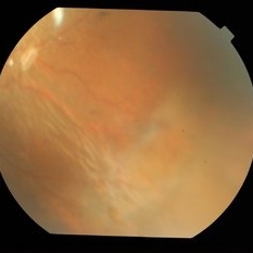
Multiple retinal folds 4 weeks following vitreous surgery (perfluorodecalin assisted) for retinal detachment with giant retinal tear.
Photographer: Mallika Goyal, MD, Apollo Health City, Jubilee Hills, Hyderabad, India
Condition/keywords: retinal fold
-
 Retinal Folds Following Retinal Reattachment Surgery
Nov 22 2015 by Mallika Goyal, MD
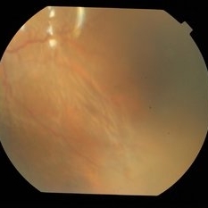
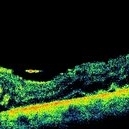
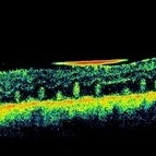
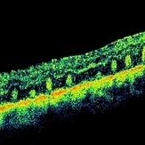
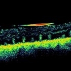
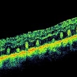
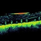
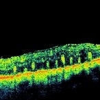
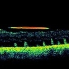
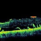
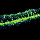
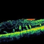
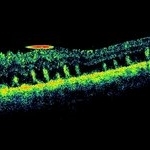
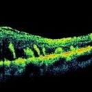
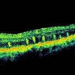
Multiple retinal folds 4 weeks following vitreous surgery (perfluorodecalin assisted) for retinal detachment with giant retinal tear. OCT shows residual subretinal fluid and outer retinal folds (ORFs) seen as vertical hyperreflective lesions consisting of folded inner segment/outer segment of photoreceptors band and external limiting membrane band.
Photographer: Mallika Goyal, MD, Apollo Health City, Jubilee Hills, Hyderabad, India
Condition/keywords: retinal fold
-
 Retinal Folds Following Retinal Reattachment Surgery
Nov 22 2015 by Mallika Goyal, MD
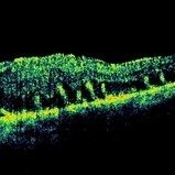
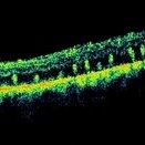
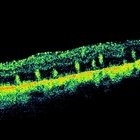
Multiple retinal folds 4 weeks following vitreous surgery (perfluorodecalin assisted) for retinal detachment with giant retinal tear. OCT shows residual subretinal fluid and outer retinal folds (ORFs) seen as vertical hyperreflective lesions consisting of folded inner segment/outer segment of photoreceptors band and external limiting membrane band.
Photographer: Mallika Goyal, MD, Apollo Health City, Jubilee Hills, Hyderabad, India
Condition/keywords: retinal fold
A project from the American Society of Retina Specialists