-
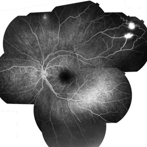
 Retinal Capillary Hemangioma
Feb 18 2016 by Hashim Ali Khan, OD, FAAO
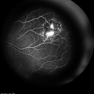
FA of 23-year-old-woman with RCH as part of spectrum of Von Hippel- Lindau
Imaging device: Heidelberg Spectralis
Condition/keywords: Von Hippel-Lindau
-
 Pattern Dystrophy
Apr 20 2016 by Hashim Ali Khan, OD, FAAO
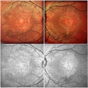
Color and NIR images of a 32-year-old woman with pattern dystrophy.
Imaging device: Spectralis Multicolor imaging
Condition/keywords: butterfly dystrophy, pattern dystrophy, pattern macular dystrophy
-
 Hemorrhagic Pigment Epithelial Detachment
Dec 14 2016 by Hashim Ali Khan, OD, FAAO
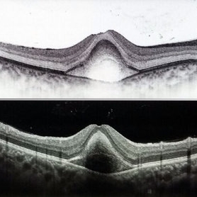
OCT of a 20-year-old female after trauma with tennis-ball, showing a hemorrhagic PED. RPE is elevated. The second Hyper-reflective band corresponding to Bruchs membrane (BM complex) is visible.
Condition/keywords: pigment epithelial detachment (PED), subretinal hemorrhage
-
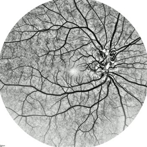
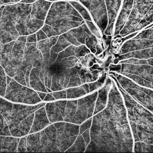
 OCTA of Diabetic Retinopathy
Mar 13 2017 by Hashim Ali Khan, OD, FAAO
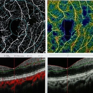
Optical coherence tomographic angiography showing capillary dropout and microaneurysms.
Imaging device: Angiovue
Condition/keywords: capillary dropouts, capillary nonperfusion, diabetic maculopathy, optical coherence tomography (OCT), retinal microaneurysms
-
 Combined Hamartoma of Retina and Retinal Pigment Epithelium
Mar 26 2018 by Hashim Ali Khan, OD, FAAO
Fundus photograph of a 12-year-old boy with combined hamartoma of retina and retinal pigment epithelium.
Condition/keywords: combined hamartoma, retinal pigment epithelium
-
 Vitreous Hemorrhage
Jun 5 2018 by Hashim Ali Khan, OD, FAAO
Resolving traumatic vitreous hemorrhage in a 50 years old male imaged with imaging slit lamp biomicroscope.
Condition/keywords: intragel hemorrhage, trauma, vitreous hemorrhage
-
 Congenital Peripapillary Vascular Loops
Jun 22 2018 by Hashim Ali Khan, OD, FAAO
Inverted FA of a 10-year-old boy with congenital peripapillary vascular loop.
Imaging device: Spectraliz
Condition/keywords: congenital prepapillary vascular loop, pediatic retina
-
 Peripapillary Vascular Loops
Jun 22 2018 by Hashim Ali Khan, OD, FAAO
FA of a 10-year-old boy with congenital peripapillary vascular loop.
Imaging device: Heidelberg Spectralis
Condition/keywords: congenital prepapillary vascular loop, pediatic retina
-
 Toxoplasma Macular Scar
Sep 22 2018 by Hashim Ali Khan, OD, FAAO
Fundus Photographs of a 17-year-old male with inactive macular toxoplasma scar.
Condition/keywords: inactive toxoplasmosis, toxoplasmosis chorioretinitis
-
 Toxoplasma Scar
Sep 22 2018 by Hashim Ali Khan, OD, FAAO
Fundus photograph of a 17-year-old male with inactive macular toxoplasma scar.
Condition/keywords: inactive toxoplasmosis, toxoplasmosis chorioretinitis
-
 Snowflake Vitreoretinal Degeneration
Nov 29 2018 by Hashim Ali Khan, OD, FAAO
Peripheral snowflake in a 16-year-old female. The fellow eye had chronic total retinal detachment.
Imaging device: Goldman triple mirror lens
Condition/keywords: peripheral fundus lesion, snowflake hereditary degeneration
-
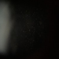 Intermediate Uveitis
Dec 14 2018 by Hashim Ali Khan, OD, FAAO
Vitreous cells in a 30-year-old female with intermediate uveitis.
Condition/keywords: vitritis
-
 Asteroid Hyalosis
Dec 16 2018 by Hashim Ali Khan, OD, FAAO
Slit-lamp photographs of a 50-year-old man with asteroid hyalosis.
Condition/keywords: asteroid hyalosis, vitreous opacity
-
 Von Hippel-Lindau Syndrome
Jan 4 2019 by Hashim Ali Khan, OD, FAAO
Montage of multiple FA images showing Retinal Capillary Hemangiomas in a 24-year-old woman with Von Hippel-Lindau syndrome.
Imaging device: Spectralis
Condition/keywords: Von Hippel-Lindau
-
 Gyrate Atrophy
Jan 6 2019 by Hashim Ali Khan, OD, FAAO
Montage of Multiple Fundus Photographs from the right eye of a 25-year-old woman with gyrate atrophy.
Photographer: Ahmed Abbass
Imaging device: Topcon TRC-NW8F
Condition/keywords: gyrate atrophy, hereditary retinal dystrophy, retinal dystrophy
-
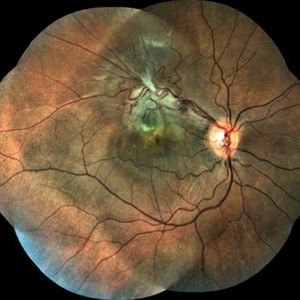
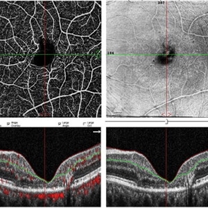 Macular Telangiectasis
May 13 2019 by Hashim Ali Khan, OD, FAAO
OCT-angio of superficial vascular network and structural OCT of a 60-years-old female demonstrating macular TEL showing alterations in FAZ and vascular remodeling and increased the intercapillary distance.
Imaging device: Optical Coherence Tomography Angiography
Condition/keywords: idiopathic macular telangiectasia, macular telangiectasia, macular telangiectasia type 2
-
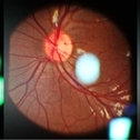
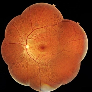
 Disc edge Veins of Kraupa
Aug 24 2019 by Hashim Ali Khan, OD, FAAO
Red free and color fundus images of 10-years-old girl with inferior disc edge veins of Kraupa; a rare exit anomaly. The inferonasal retina is drained through the venous trunk exiting at the edge of optic disc.
Condition/keywords: disc edge veins of Kraupa, vascular exit anomalies
-
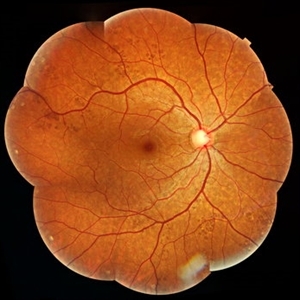
 Disc Edge Veins of Kraupa
Aug 24 2019 by Hashim Ali Khan, OD, FAAO
Red free and color fundus images of 10-year-old girl with inferior disc edge veins of Kraupa; a rare exit anomaly. The inferonasal retina is drained through the venous trunk exiting at the edge of optic disc.
Condition/keywords: disc edge veins of Kraupa, vascular exit anomalies
-
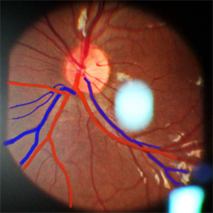 Disc Edge Veins of Kraupa
Aug 25 2019 by Hashim Ali Khan, OD, FAAO
Color fundus with overlay drawing of 10-year-old girl with inferior disc edge veins of Kraupa; a rare exit anomaly. The inferonasal retina is drained through the venous trunk exiting at the edge of optic disc.
Condition/keywords: disc edge veins of Kraupa, vascular exit anomalies
-
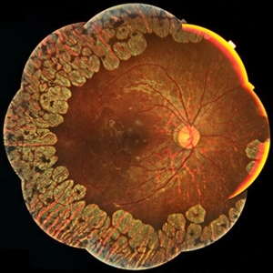
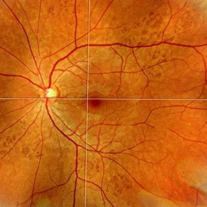 Congenital Hypertrophy of Retinal Pigment Epithelium (CHRPE)
Sep 6 2019 by Hashim Ali Khan, OD, FAAO
Color fundus montage of a 22-year-old man with congenital hypertrophy of retinal pigment epithelium.
Imaging device: TOPCON TRC NW8F
Condition/keywords: bear tracks, congenital hypertrophy of the retinal pigment epithelium (CHRPE), RPEH-FAP
-
 Congenital Hypertrophy of Retinal Pigment Epithelium (CHRPE)
Sep 7 2019 by Hashim Ali Khan, OD, FAAO
Color fundus montage of a 22-year-old man with congenital hypertrophy of retinal pigment epithelium..
Condition/keywords: bear tracks, congenital hypertrophy of the retinal pigment epithelium (CHRPE)
-
 Congenital Hypertrophy of Retinal Pigment Epithelium
Sep 7 2019 by Hashim Ali Khan, OD, FAAO
Color fundus montage of a 22-year-old man with congenital hypertrophy of retinal pigment epithelium.
Condition/keywords: bear tracks, congenital hypertrophy of the retinal pigment epithelium (CHRPE), RPE hyperplasia
-
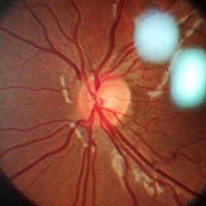 Disc Edge Veins of Kraupa
Sep 15 2019 by Hashim Ali Khan, OD, FAAO
Color fundus with overlay drawing of 12-year-old girl with superonasal disc edge veins of Kraupa; a rare exit anomaly. The superonasal retina is drained through the venous trunk exiting at the edge of optic disc.
Condition/keywords: disc edge veins of Kraupa, vascular exit anomalies
-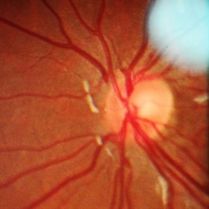
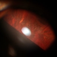
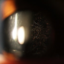 Schaffer's Sign
Dec 23 2019 by Hashim Ali Khan, OD, FAAO
Brown iris pigment in vitreous of a pseudophakic eye without retinal detachment or breaks/ holes in retina.
Condition/keywords: detached vitreous, Schaffer's sign, vitreous pigment
A project from the American Society of Retina Specialists