-
 Combined Hamartoma of Retina and Retinal Pigment Epithelium
Apr 30 2021 by ARVIND JAIN M
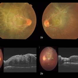
A 26-year-old gentlemen came with complains of defective vision in both eyes since childhood. His BCVA was right eye 5/60 and left eye 6/60. His anterior segment examination showed no abnormality with posterior segment examination showed both eyes (1a and 1b) greyish white elevated lesion involving the macula with thick fibrotic epiretinal membrane causing the macular drag temporally in right eye and supero-temporally in left eye. (2a and 2b) showing the thick ERM with the hamartoma of the retina and RPE.
Photographer: DR ARVIND JAIN, ARAVIND EYE HOSPITAL, COIMBATORE,INDIA
Condition/keywords: combined hamartoma, congenital hypertrophy of the retinal pigment epithelium (CHRPE), epiretinal membrane (ERM), retinal pigment epithelium (RPE) hamartoma
-
 Congenital Hamartoma of Retina and RPE
May 23 2024 by ARVIND JAIN M
Bilateral involving CHRRPE lesion in a 26 year old gentleman who came with complains of defective vision in both eyes since childhood. His BCVA was Right eye 5/60 and left eye 6/60. His anterior segment examination showed no abnormality with posterior segment examination showed both eyes (Fig.1a and 1b) greyish white elevated lesion involving the macula with thick fibrotic membrane causing the macular drag temporally in right eye and supero-temporally in left eye. (Fig.2a and 2b) OCT showing the thick ERM with the disorganized inner retinal layers suggestive of hamartoma of the Retina and Retinal Pigment Epithelium.
Photographer: Dr. Arvind Jain M, MBBS,MS Ophthal, FVRS
Condition/keywords: CHRRPE
-
 Coats Disease
May 23 2024 by ARVIND JAIN M
a.right eye fundus image and b. FFA montage of a 8 year old boy showing light bulb aneurysms of the arterioles with exudation with sub retinal fibrosis and telangiectasia in periphery who complained of defective vision, classical of coats disease.
Photographer: Dr. Arvind Jain M, MBBS,MS Ophthal, FVRS
Condition/keywords: COATS DISEASE, Leber's miliary aneurysm, light-bulb aneurysms
-
Tractional RD-Making the Decision When and Where to Stop
May 23 2024 by ARVIND JAIN M
This is a young gentlemen with defective vision for 3 months in his right eye. He gave the history of recurrent redness of the right past few months. he was diagnosed to have right eye vasculitis with tractional detachment. He underwent uveitic workup and under steroid cover right eye paraplana vitrectomy with membrane peeling with endolaser with c3f8 gas was planned. patient improved significantly. this surgical video demonstrates when and where to stop during membrane peeling and get good results.
Condition/keywords: Eales disease, retinal vasculitis, tractional retinal detachment
A project from the American Society of Retina Specialists