An 83 year-old woman was referred with mild painless progressive blurring of central vision OU over the past several months. She was noted to have visual acuities of 20/200 OD and 20/30 OS. Macular exam revealed symmetric yellow subfoveal pigment clumping. Angiography revealed early blockage and some mild late staining due to the pigment clumping without any evidence of choroidal neovascularization. OCT revealed typical hyper-reflective subretinal elevation. This disorder typically begins in middle age, presenting with symmetric foveal pigment clumping and minimal vision loss. Atrophic changes can develop later. It has been given multiple names over time including adult foveomacular vitelliform dysrophy, adult onset vitelliform macular dystrophy, adult foveal pigment epithelial dystrophy, adult onset foveal macular dystrophy, etc. It is caused by mutations in the PRPH2 or BEST1 genes and usually inherited in an autosomal dominant fashion, although not all individuals who inherit the mutated gene develop symptoms.
-
 Adult Foveomacular Vitelliform Dystrophy
Dec 27 2014 by Thomas A. Ciulla, MD, MBA, FASRS
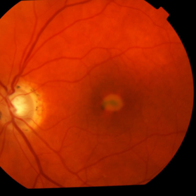
An 83-year-old woman was referred with mild painless progressive blurring of central vision OU over the past several months. She was noted to have visual acuities of 20/200 OD and 20/30 OS. Macular exam revealed symmetric yellow subfoveal pigment clumping.
Photographer: Thomas Steele
Condition/keywords: adult foveomacular dystrophy, adult vitelliform dystrophy
-
 Adult Foveomacular Vitelliform Dystrophy
Dec 27 2014 by Thomas A. Ciulla, MD, MBA, FASRS
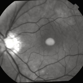
An 83-year-old woman was referred with mild painless progressive blurring of central vision OU over the past several months. She was noted to have visual acuities of 20/200 OD and 20/30 OS. Macular exam revealed symmetric yellow subfoveal pigment clumping.
Photographer: Thomas Steele
Condition/keywords: adult foveomacular dystrophy, adult vitelliform dystrophy
-
 Adult Foveomacular Vitelliform Dystrophy
Dec 27 2014 by Thomas A. Ciulla, MD, MBA, FASRS
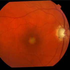
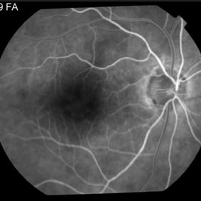
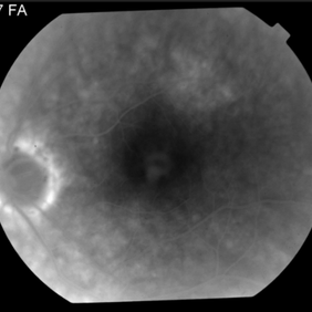
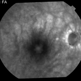
Angiography revealed early blockage and some mild late staining due to the pigment clumping without any evidence of choroidal neovascularization.
Photographer: Thomas Steele
Condition/keywords: adult foveomacular dystrophy, adult vitelliform dystrophy
-
 Adult Foveomacular Vitelliform Dystrophy
Dec 27 2014 by Thomas A. Ciulla, MD, MBA, FASRS
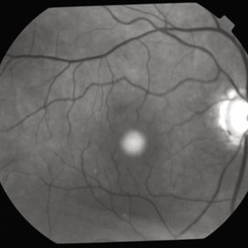
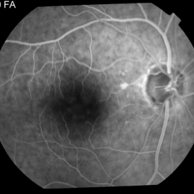
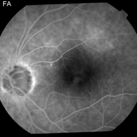
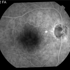
OCT revealed typical hyper-reflective subretinal elevation.
Condition/keywords: adult foveomacular dystrophy, adult vitelliform dystrophy