-
 Heredomacular Degeneration
May 4 2014 by Mallika Goyal, MD
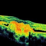
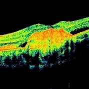
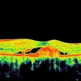
OCT left eye of a 16-year-old boy with bilateral heredomacular degeneration shows development of a large central scar 18 months after presentation subsequent to 3 intravitreal avastin injections, with slight improved visual acuity from 20/40 at presentation to 20/25 subsequently. Right eye has remained stable with good visual acuity and no intervention.
Photographer: Mallika Goyal, MD, Apollo Health City, Jubilee Hills, Hyderabad, India
Condition/keywords: heredomacular degeneration
-
 Heredomacular Degeneration
May 4 2014 by Mallika Goyal, MD
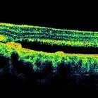
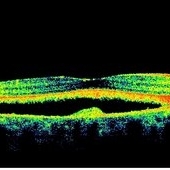
OCT scan inferior to foveal centre shows cavity statusidentical to that at presentation 18 months prior to this scan.
Photographer: Mallika Goyal, MD, Apollo Health City, Jubilee Hills, Hyderabad, India
Condition/keywords: heredomacular degeneration, optical coherence tomography (OCT)
-
 Heredomacular Degeneration
May 4 2014 by Mallika Goyal, MD
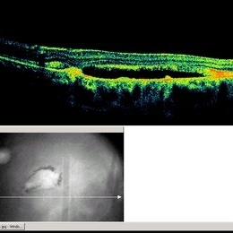
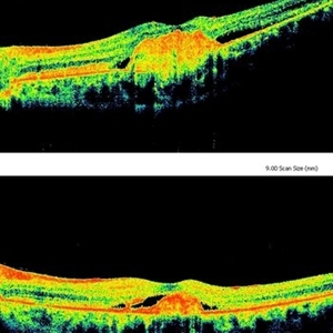
Change in OCT status over 6 months of intravitreal avastin in a 14-year-old male child with bilateral heredomacular degeneration. The lower figure is status at presentation and the upper one 6 months later. OCT and clinical exam reveal enlargement of central scar with slight improved visual acuity from 20/40 at presentation to 20/25 subsequently.
Photographer: Mallika Goyal, MD, Apollo Health City, Jubilee Hills, Hyderabad, India
Condition/keywords: heredomacular degeneration, optical coherence tomography (OCT)
-
 Heredomacular Degeneration
May 3 2014 by Mallika Goyal, MD
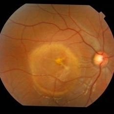
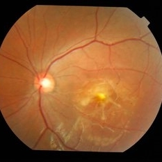
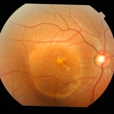
Right fundus of a 15-year-old boy with bilateral heredomacular degeneration.
Photographer: Mallika Goyal, MD, Apollo Health City, Jubilee Hills, Hyderabad, India
Condition/keywords: heredomacular degeneration
-
 Heredomacular Degeneration
May 3 2014 by Mallika Goyal, MD
Left fundus of a 15-year-old boy with bilateral heredomacular degeneration. Lesion has become circumscribed with a central scar developing over the 8 months he been under evaluation (and management with 3 injections of avastin at 3 monthly intervals at presentation). Visual acuity 20/25,
Photographer: Mallika Goyal, MD, Apollo Health City, Jubilee Hills, Hyderabad, India
Condition/keywords: heredomacular degeneration
-
---thumb.JPG/image-square;max$300,300.ImageHandler) Heredomacular Degeneration
Heredomacular Degeneration
Nov 18 2013 by Mallika Goyal, MD
Fluorescein angiogram of right eye of a 14-year-old boy with heredomacular degeneration.
Photographer: Mallika Goyal, MD, Apollo Health City, Hyderabad
Condition/keywords: heredomacular degeneration
-
---thumb.JPG/image-square;max$300,300.ImageHandler) Heredomacular Degeneration
Heredomacular Degeneration
Nov 18 2013 by Mallika Goyal, MD
Late phase fluorescein angiogram of right eye of a 14-year-old boy with heredomacular degeneration.
Photographer: Mallika Goyal, MD, Apollo Health City, Hyderabad
Condition/keywords: heredomacular degeneration
-
---thumb.JPG/image-square;max$300,300.ImageHandler) Heredomacular Degeneration
Heredomacular Degeneration
Nov 18 2013 by Mallika Goyal, MD
Fluorescein angiogram of left eye of a 14-year-old boy with heredomacular degeneration.
Photographer: Mallika Goyal, MD, Apollo Health City, Hyderabad
Condition/keywords: heredomacular degeneration
-
---thumb.JPG/image-square;max$300,300.ImageHandler) Heredomacular Degeneration
Heredomacular Degeneration
Nov 18 2013 by Mallika Goyal, MD
Late phase fluorescein angiogram of left eye of a 14-year-old boy with heredomacular degeneration.
Photographer: Mallika Goyal, MD, Apollo Health City, Hyderabad
Condition/keywords: heredomacular degeneration
-
 Heredomacular Degeneration
Dec 7 2013 by Mallika Goyal, MD
OCT of the left eye of a young boy with bilateral heredomacular degeneration with cavitation and OCT-dense deposits at macula.
Photographer: Mallika Goyal, MD, Apollo Health City, Jubilee Hills, Hyderabad
Condition/keywords: heredomacular degeneration, optical coherence tomography (OCT)
-
 Heredomacular Degeneration
Dec 7 2013 by Mallika Goyal, MD
OCT of the right eye of a young boy with bilateral heredomacular degeneration shows large cavity (from splitting of the RPE) and exudate at macula.
Photographer: Mallika Goyal, MD, Apollo Health City, Jubilee Hills, Hyderabad
Condition/keywords: heredomacular degeneration, optical coherence tomography (OCT)
-
 Heredomacular Degeneration
Dec 7 2013 by Mallika Goyal, MD
OCT of the left eye of a young boy with bilateral heredomacular degeneration with cavitation and OCT-dense deposits at macula.
Photographer: Mallika Goyal, MD, Apollo Health City, Hyderabad, India
Condition/keywords: heredomacular degeneration, optical coherence tomography (OCT)
-
 Heredomacular Degeneration
May 3 2014 by Mallika Goyal, MD
Right fundus of a 15-year-old boy with bilateral heredomacular degeneration.
Photographer: Mallika Goyal, MD, Apollo Health City, Jubilee Hills, Hyderabad, India
Condition/keywords: heredomacular degeneration
-
---thumb.JPG/image-square;max$300,300.ImageHandler) Heredomacular Degeneration
Heredomacular Degeneration
Nov 18 2013 by Mallika Goyal, MD
Left fundus of a 14-year-old boy with bilateral heredomacular degeneration. Visual acuity 20/100. No consanguinity or similar family history.
Photographer: Mallika Goyal, MD, Apollo Health City, Hyderabad
Condition/keywords: heredomacular degeneration
-
 Heredomacular Degeneration
May 3 2014 by Mallika Goyal, MD
Right fundus of a 15-year-old boy with bilateral heredomacular degeneration.
Photographer: Mallika Goyal, MD, Apollo Health City, Jubilee Hills, Hyderabad, India
Condition/keywords: heredomacular degeneration
-
 Heredomacular Degeneration
May 3 2014 by Mallika Goyal, MD
Left fundus of a 15-year-old boy with bilateral heredomacular degeneration. Lesion has become circumscribed with a central scar developing over the 14 months he been under evaluation (and management with 3 injections of avastin at 3 monthly intervals at presentation). Visual acuity 20/30,
Photographer: Mallika Goyal, MD, Apollo Health City, Jubilee Hills, Hyderabad, India
Condition/keywords: heredomacular degeneration
-
---thumb.JPG/image-square;max$300,300.ImageHandler) Heredomacular Degeneration
Heredomacular Degeneration
Nov 18 2013 by Mallika Goyal, MD
Right fundus of a 14-year-old boy with bilateral heredomacular degeneration. Visual acuity 20/20, unaffected this eye. No consanguinity or similar family history.
Photographer: Mallika Goyal, MD, Apollo Health City, Hyderabad
Condition/keywords: heredomacular degeneration
-
 Heredomacular Degeneration
May 3 2014 by Mallika Goyal, MD
Left fundus of a 15-year-old boy with bilateral heredomacular degeneration. Lesion has become circumscribed with a prominent central scar over the 18 months he been under evaluation (and management with 3 injections of avastin at 3 monthly intervals at presentation). Visual acuity 20/25,
Photographer: Mallika Goyal, MD, Apollo Health City, Jubilee Hills, Hyderabad, India
Condition/keywords: heredomacular degeneration
-
 Heredomacular Degeneration
May 3 2014 by Mallika Goyal, MD
Left fundus of a 15-year-old boy with bilateral heredomacular degeneration. Lesion has become circumscribed with a central scar developing over the 10 months he been under evaluation (and management with 3 injections of avastin at 3 monthly intervals at presentation). Visual acuity 20/30,
Photographer: Mallika Goyal, MD, Apollo Health City, Jubilee Hills, Hyderabad, India
Condition/keywords: heredomacular degeneration
A project from the American Society of Retina Specialists