-
 Torpedo Maculopathy
Jan 20 2020 by Pierre-Henry Gabrielle, MD
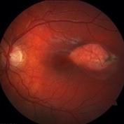
Fundus photograph of an asymptomatic 12-year-old girl with torpedo maculopathy of the left eye.
Photographer: Pierre-Henry Gabrielle, Ophthalmology department, Dijon University Hospital, France
Imaging device: Zeiss visucam
Condition/keywords: fundus photograph, torpedo maculopathy
-
 Torpedo Maculopathy
Jan 20 2020 by Pierre-Henry Gabrielle, MD
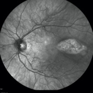
IR imaging photograph of an asymptomatic 12-year-old girl with torpedo maculopathy of the left eye.
Photographer: Pierre-Henry Gabrielle, Ophthalmology department, Dijon University Hospital, France
Imaging device: Heidelberg Spectralis
Condition/keywords: infrared image, torpedo maculopathy
-
 Torpedo Maculopathy
Jan 20 2020 by Pierre-Henry Gabrielle, MD
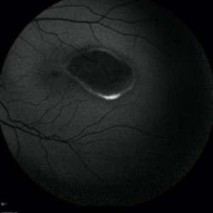
Fundus autofluorescence imaging of an asymptomatic 12-year-old girl with torpedo maculopathy of the left eye.
Photographer: Pierre-Henry Gabrielle, Ophthalmology department, Dijon University Hospital, France
Imaging device: Heidelberg Spectralis
Condition/keywords: fundus autofluorescence (FAF), torpedo maculopathy
-
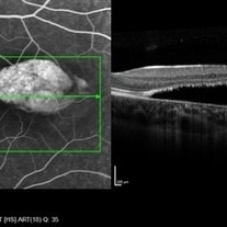
 Torpedo Maculopathy
Jan 20 2020 by Pierre-Henry Gabrielle, MD
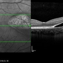
Coupled OCT B-scan and IR imaging of an asymptomatic 12-year-old girl with torpedo maculopathy of the left eye. One can see that there is no involvement of the fovea which explain that the patient has no visual sign.
Photographer: Pierre-Henry Gabrielle, Ophthalmology department, Dijon University Hospital, France
Imaging device: Heidelberg Spectralis
Condition/keywords: optical coherence tomography (OCT), torpedo maculopathy
-
 Torpedo Maculopathy
Jan 20 2020 by Pierre-Henry Gabrielle, MD
Coupled OCT B-scan and fluorescein angiogram of an asymptomatic 12-year-old girl with torpedo maculopathy of the left eye. One can report complete RPE atrophy at lesion site with window defect on FA and choroidal cavitation on OCT.
Photographer: Pierre-Henry Gabrielle, Ophthalmology department, Dijon University Hospital, France
Imaging device: Heidelberg Spectralis
Condition/keywords: fluorescein angiogram (FA), optical coherence tomography (OCT), torpedo maculopathy
-
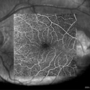 Torpedo Maculopathy
Jan 20 2020 by Pierre-Henry Gabrielle, MD
Superficial capillary plexus OCT-angiography of an asymptomatic 12-year-old girl with Torpedo maculopathy of the left eye. No abnormality.
Photographer: Pierre-Henry Gabrielle, Ophthalmology department, Dijon University Hospital, France
Imaging device: Zeiss Cirrus OCT
Condition/keywords: optical coherence tomography (OCT), torpedo maculopathy
-
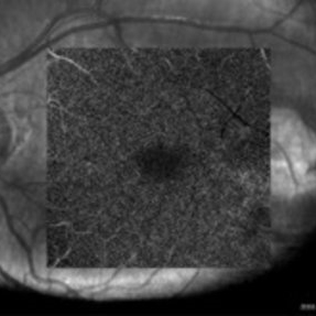 Torpedo Maculopathy
Jan 20 2020 by Pierre-Henry Gabrielle, MD
Deep capillary plexus OCT-angiography of an asymptomatic 12-year-old girl with torpedo maculopathy of the left eye. No abnormality.
Photographer: Pierre-Henry Gabrielle, Ophthalmology department, Dijon University Hospital, France
Imaging device: Zeiss Cirrus OCT
Condition/keywords: optical coherence tomography (OCT), torpedo maculopathy
-
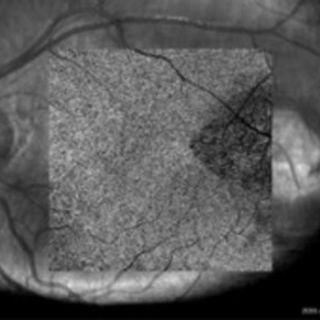 Torpedo Maculopathy
Jan 20 2020 by Pierre-Henry Gabrielle, MD
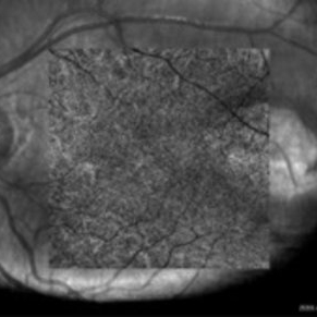
Choriocapillaris OCT-angiography of an asymptomatic 12-years-old girl with Torpedo maculopathy of the left eye. One can see a diffuse flow attenuation at choriocapillaris segmentation level at the lesion site.
Photographer: Pierre-Henry Gabrielle, Ophthalmology department, Dijon University Hospital, France
Imaging device: Zeiss Cirrus OCT
Condition/keywords: optical coherence tomography (OCT), torpedo maculopathy
-
 Torpedo Maculopathy
Jan 20 2020 by Pierre-Henry Gabrielle, MD
Choroidal vessels OCT-angiography of an asymptomatic 12-year-old girl with Torpedo maculopathy of the left eye. No abnormality.
Photographer: Pierre-Henry Gabrielle, Ophthalmology department, Dijon University Hospital, France
Imaging device: Zeiss Cirrus OCT
Condition/keywords: optical coherence tomography (OCT), torpedo maculopathy
A project from the American Society of Retina Specialists