 Initializing download.
-
 By Niloofar Piri, MD
SSM Health Group, St Louis University
Co-author(s): Jennifer Sim, MD, Saint Louis University ; Anahita Dadali, MBBS student, St George's, University of London - Uploaded on Jul 22, 2021.
- Last modified by Caroline Bozell on Jul 22, 2021.
- Rating
- Appears in
- Miscellaneous
- Condition/keywords
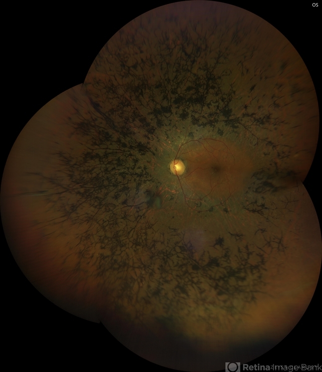
- retinitis pigmentosa
- Photographer
- Niloofar Piri, MD
- Imaging device
- Fundus camera
- Description
- Montage wide field fundus photograph of the left eye of the same patient. 55-year-old male presented with late onset night blindness and peripheral vision loss for one year. Central vision is preserved at 20/25. ERG demonstrated extinguished rod function, and minimally diminished cone function. Waxy pallor of the optic nerve, arterial narrowing, and peripheral bony spicules are the classic triad of RP which are demonstrated in the photograph.


---thumb.jpg/image-square;max$79,0.ImageHandler "Retinitis Pigmentosa")


