 Initializing download.
-
 By John S. King, MD
Retina Associates, PA
Co-author(s): Kent Zocchi, MD - Uploaded on Jun 1, 2019.
- Last modified by Joshua Friedman on Feb 23, 2022.
- Rating
- Appears in
- Presumptive FAME
- Condition/keywords
- macular edema, Gilenya, cystoid macular edema (CME)
- Photographer
- Kay Dalby
- Imaging device
-
Fundus camera
Topcon 50 - Description
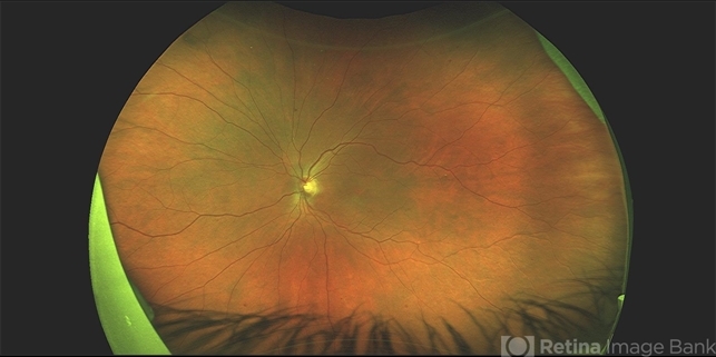
- 60 year old Caucasian female with two week history of decreased vision in the left eye. Background history includes multiple sclerosis for which she uses Gilenya for the past five years, and no history of uveitis or recent MS relapse. Her vision in the left eye was 20/100 J5. The eye appeared overall "quiet" with the exception of rare cells in the anterior vitreous. The fundus appearance and FA can be seen in the images provided. The fundus image shows a blunt foveal reflex , and a few mid-peripheral IRHs, OS only. Retinal hemorrhages have been reported in a few articles as possibly associated with this medication, but the IRHs here are much less prominent than the ones reported (see below). STK was administered and neurologist was able to discontinue the Gilenya. 10 days later the CME had decreased significantly. 8 weeks later the edema had resolved as seen in the OCT images of the initial and latest appearance (the IRHs have decreased as well) Bhatti MT, Freedman SM, Mahmoud TH. Fingolimod therapy and macular haemorrhage. J Neuroophthalmol 2013; 33(4): 370–372. Ueda N, Saida K. Retinal haemorrhages following fingolimod treatment for multiple sclerosis; a case report. BMC Ophthalmol 2015; 15(1): 135.

")
")
")
")
")
")


