 Initializing download.
-
 By John S. King, MD
Retina Associates, PA
Co-author(s): Paul Hruby, MD - Uploaded on Oct 23, 2018.
- Last modified by Caroline Bozell on Oct 23, 2018.
- Rating
- Appears in
- Purtscher Retinopathy
- Condition/keywords
- Purtscher's retinopathy, cystoid macular edema (CME), acute pancreatitis
- Imaging device
-
Fundus camera
Optos CA - Description
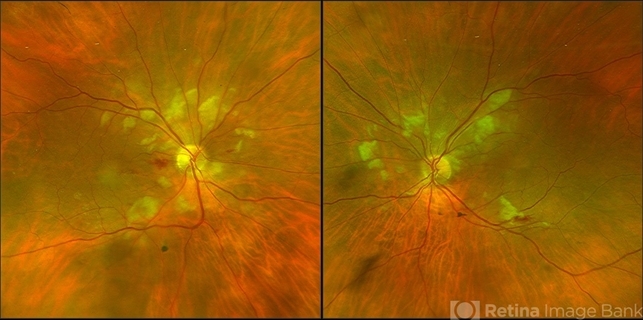
- 70-year-old white female with with recent admission to hospital for acute pancreatitis, developed acute decrease in vision while in the hospital. Was seen by Dr Hruby in the outpatient clinic within a week of hospitalization. Visual acuity sc was 20/150 OU ph to 20/100 in each eye. Posterior segment showed Purtscher flecken, a few superficial IRHs nasal to the optic nerve, and CWSs. OCT revealed macular thickening with both CME and SRF. The FA did not show any significant CME. There was blockage in the areas of the Purtscher flecken (see image) with some staining of the vessels contiguous to them; possible disc leakage. Patient was monitored and each month vision and OCT improved. On the last visit, visual acuity sc was 20/40 OD ph to 20/25, and 20/30 OS without improvement with ph. The retinas and OCT had normalized, and patient has noticed marked improvement.
")
")
")
")
")
")


