 Initializing download.
-
 By John S. King, MD
Retina Associates, PA - Uploaded on Jan 7, 2018.
- Last modified by Chayal Patel on Jan 16, 2018.
- Rating
- Appears in
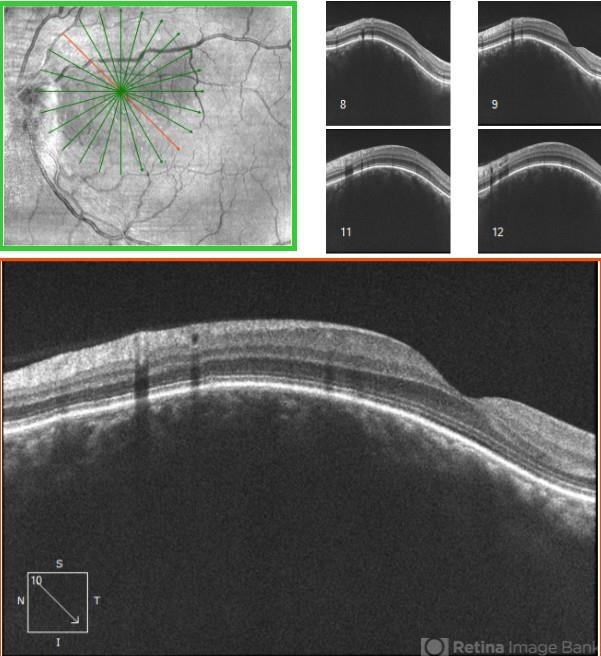
- Discrete Choroidal Hemangioma
- Condition/keywords
- choroidal hemangioma
- Imaging device
-
Optical coherence tomography system
Cirrus - Description
- 49-year-old WF seen for annual, routine exam, and sent here because could not refract to better than 20/40. Optos Photos and FA showed minimal to no findings, while topcon photos showed a well curcumscribed red/orange lesion with high internal reflectivity on Bscan, and sparing of choriocapillaris on OCT; no SRF, exudates, or other findings present.


