Search results (0 results)
-
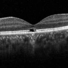
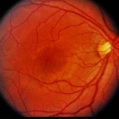
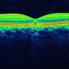
 Solar Retinopathy With IS-OS Focal Defects
Dec 18 2025 by Deepak Bhojwani, MS
Color fundus image and oct of a healthy 23 year old male complaining of blurry vision following watching an eclipse with naked eyes during his childhood days. Fundus image shows focal pigmentary changes on fovea and oct shows IS-OS defects with disruption of RPE (focal changes in outer retinal layers) suggestive of solar retinopathy.
Photographer: DR DEEPAK BHOJWANI
Imaging device: FUNDUS CAMERA
Condition/keywords: outer retina defects, solar retinopathy
-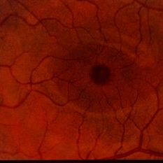
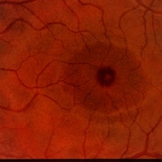
 Solar Retinopathy
Dec 18 2025 by Deepak Bhojwani, MS
Color fundus image and oct of a healthy 23 year old male complaining of blurry vision following watching an eclipse with naked eyes during his childhood days. Fundus image shows focal pigmentary changes on fovea and OCT shows is-os defects with disruption of RPE (focal changes in outer retinal layers) suggestive of solar retinopathy.
Photographer: DR DEEPAK BHOJWANI
Imaging device: FUNDUS CAMERA
Condition/keywords: outer retina defects, solar retinopathy
-
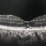
 Solar Retinopathy
Apr 1 2025 by Isaac Agranoff
OCT scan of 18-year-old male presenting with 20/40 BCVA OU and bilateral focal outer retinal subfoveal defects. Patient reported long-term history of frequent sungazing, has stopped within past 6-9 months.
Photographer: Isaac Agranoff
Imaging device: Heidelberg Spectralis
Condition/keywords: solar retinopathy
-
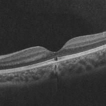
 Solar Retinopathy
Mar 17 2024 by Hector Gabriel Moreno Solano, MD, MHA
OCT scan of a 65 year old male with a history of direct exposure to solar eclipse rays, visual acuity of affected eye 20/80, contralateral eye 20/25.
Photographer: Héctor Gabriel Moreno-Solano, MD, MHA
Imaging device: Revo Optopol
Condition/keywords: light toxicity, macula, solar retinopathy
-
 Solar Retinopathy
Jun 27 2020 by Thirumalesh Mochi Basavaraj, MD
Fundus photo showing eclipse burn, Inset OCT picture shows loss of photoreceptor layer.
Photographer: Ravikrishna, Puttaswamy
Imaging device: Heidelberg Spectralis
Condition/keywords: solar retinopathy
-
 Solar Retinopathy
-
 Solar Retinopathy
-
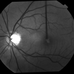
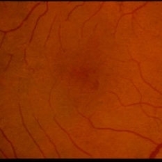
 Solar Retinopathy
Sep 18 2015 by David Callanan, MD
22-year-old male, solar retinopathy.
Condition/keywords: solar retinopathy
-
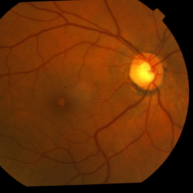
 Solar Retinopathy
Sep 18 2015 by David Callanan, MD
22-year-old male, solar retinopathy.
Condition/keywords: solar retinopathy
-
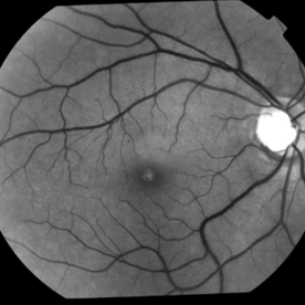
 Solar Retinopathy
Sep 18 2015 by David Callanan, MD
22-year-old male, solar retinopathy.
Condition/keywords: solar retinopathy
-
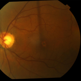
 Solar Retinopathy
Sep 18 2015 by David Callanan, MD
22-year-old male, solar retinopathy.
Condition/keywords: solar retinopathy
-
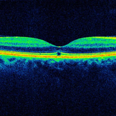
 Solar Maculopathy, OCT, Left Macula
Mar 7 2015 by Thomas A. Ciulla, MD, MBA, FASRS
OCT revealed symmetric focal discontinuity of the IS/OS line and underlying RPE.
Condition/keywords: solar maculopathy, solar retinopathy
-
 Solar Maculopathy, OCT, Right Macula
Mar 7 2015 by Thomas A. Ciulla, MD, MBA, FASRS
OCT revealed symmetric focal discontinuity of the IS/OS line and underlying RPE.
Condition/keywords: solar maculopathy, solar retinopathy
-
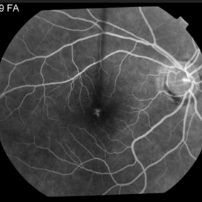
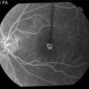
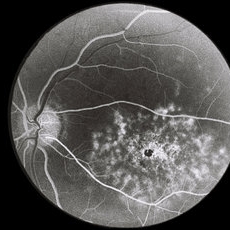
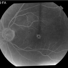 Solar Maculopathy
Mar 7 2015 by Thomas A. Ciulla, MD, MBA, FASRS
Angiography revealed staining centrally OD and blockage by the central pigment clumping with staining of surrounding atrophy OS only.
Photographer: Thomas Steele
Condition/keywords: solar maculopathy, solar retinopathy
-
 Solar Maculopathy
Mar 7 2015 by Thomas A. Ciulla, MD, MBA, FASRS
Angiography revealed staining centrally OD and blockage by the central pigment clumping with staining of surrounding atrophy OS only.
Photographer: Thomas Steele
Condition/keywords: solar maculopathy, solar retinopathy
-
 Solar Maculopathy
Mar 7 2015 by Thomas A. Ciulla, MD, MBA, FASRS
Angiography revealed staining centrally OD and blockage by the central pigment clumping with staining of surrounding atrophy OS only.
Photographer: Thomas Steele
Condition/keywords: solar maculopathy, solar retinopathy
-
 Solar Maculopathy
Mar 7 2015 by Thomas A. Ciulla, MD, MBA, FASRS
Angiography revealed staining centrally OD and blockage by the central pigment clumping with staining of surrounding atrophy OS only.
Photographer: Thomas Steele
Condition/keywords: solar maculopathy, solar retinopathy
-
 Solar Maculopathy
Mar 7 2015 by Thomas A. Ciulla, MD, MBA, FASRS
On the left, macular examination revealed central pigment clumping with surrounding atrophy.
Photographer: Thomas Steele
Condition/keywords: solar maculopathy, solar retinopathy
-
 Solar Maculopathy
Mar 7 2015 by Thomas A. Ciulla, MD, MBA, FASRS
Macular examination revealed mild atrophy central on the right.
Photographer: Thomas Steele
Condition/keywords: solar maculopathy, solar retinopathy
-
 Solar Maculopathy
Mar 7 2015 by Thomas A. Ciulla, MD, MBA, FASRS
Macular examination revealed mild atrophy central on the right.
Photographer: Thomas Steele
Condition/keywords: solar maculopathy, solar retinopathy
-
 Solar Maculopathy
Mar 7 2015 by Thomas A. Ciulla, MD, MBA, FASRS
On the left, macular examination revealed central pigment clumping with surrounding atrophy.
Photographer: Thomas Steele
Condition/keywords: solar maculopathy, solar retinopathy
-
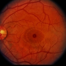
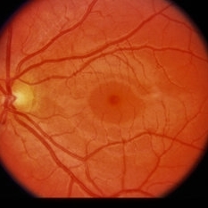 Solar Retinopathy
Feb 12 2015 by H. Michael Lambert, MD
Yellow white spot on central fovea, serous detachment on macular.
Condition/keywords: macular, solar retinopathy
-
 Solar Retinopathy
Feb 12 2015 by H. Michael Lambert, MD
Yellow white spot on central fovea, serous detachment on macular.
Condition/keywords: macular, solar retinopathy
-
 Solar Retinopathy
 Loading…