Search results (14 results)
-
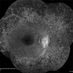
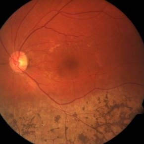
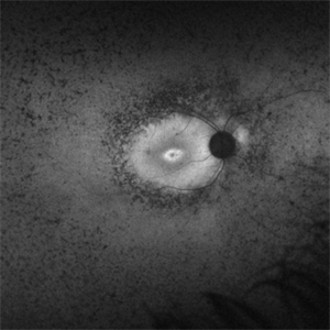 Retinitis Pigmentosa Bullseye Appearing Autofluorescence
Feb 4 2025 by Isaac Agranoff
Fundus Autofluorescence of a 14-year-old boy with suspected RP. ERG performed afterwards was almost flat. VA measured at 20/30 but with extensive constriction of confrontational visual fields. Currently awaiting genetic testing.
Photographer: Isaac Agranoff
Imaging device: Optos California
Condition/keywords: fundus autofluorescence (FAF), retinitis pigmentosa, RP
-
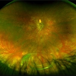
 Retinitis Pigmentosa with PPRPE
Jan 27 2025 by Vishal Agrawal, MD, FRCS,FACS,FASRS
16 year-old male patient presented with DOV, nyctalopia and nystagmus. Fundus revealed pigment clumping, pale disc and preserved para-arteriolar retinal pigment epithelium (PPRPE) in both eyes. Genetic testing revealed CRB1 gene mutation.
Photographer: Dr Ayushi
Imaging device: Clarus 700
Condition/keywords: retinitis pigmentosa
-
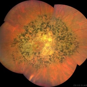
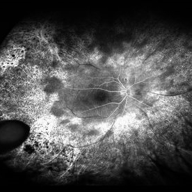
 Pericentral Retinitis Pigmentosa
Sep 6 2024 by Mauricio Bayram-Suverza, MD
A 65-year-old male patient reports experiencing bilateral blind spots that have gradually intensified over time. Genetic testing was unrevealing. The fundus autofluorescence image shows a hypoautofluorescent ring in the posterior pole, especially nasal to the nerve and along arcades.
Photographer: Mauricio Bayram-Suverza, Casey Eye Institute, OHSU.
Imaging device: Optos California
Condition/keywords: fundus autofluorescence (FAF), inherited retinal disease, nyctalopia, retinal dystrophy, retinitis pigmentosa
-
 Dislocated Lens
Apr 26 2023 by Chloe Hanifan
Ultra wide field fundus photograph of a 41-year-old male with a dislocated lens affecting his right eye. IOL noted inferior vitreous base and vitrectomy surgery for removal of IOL was recommended. Patient has history of retinitis pigmentosa as well. Patient's vision at the time of presentation was counting fingers at 2 feet.
Photographer: Chloe Hanifan
Imaging device: Optos California
Condition/keywords: dislocated lens, fundus photography, Optos, pseudocolor, retinitis pigmentosa, ULTRA WIDE FIELD
-
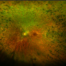
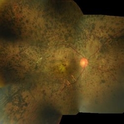
 Tapetoretinal Degeneration
Sep 7 2022 by JEFFERSON R SOUSA, Tecg.º (Biomedical Systems Technology)
Patient 52 years old, Male, progressive loss of vision since the age of 20. Retinography showed mobilization of pigments in osteoblasts, extensive area of atrophy of the pigmentary epithelium and choroid. On fluorescein angiography, typical changes following the characteristic patterns of paracentra retinal retinitis pigmentosa. Autofluorescent fundus with a sectorial autohypofluorescence pattern in the regions of atrophies.
Photographer: JEFFERSON ROCHA DE SOUSA - Retinal Department at Instituto Dr. Suel Abujamra Sao Paulo-Brazil
Imaging device: Clarus 700 - Zeiss, composite of four 135 degree images.
Condition/keywords: pericentral retinitis pigmentosa, tapeoretinal degeneration
-
 Retinitis Pigmentosa With Cystoid Macular Edema
Jun 24 2018 by Zachary M Bodnar, MD
64-year-old female with retinitis pigmentosa and cystoid macular edema, both eyes.
Imaging device: optos
Condition/keywords: cystoid macular edema (CME), retinitis pigmentosa
-
 Retinitis Pigmentosa
Aug 8 2017 by Ginny Martinez
F/A montage of a 24-year-old male with retinitis pigmentosa.
Photographer: Ginny Martinez
Condition/keywords: retinitis pigmentosa
-
 RP Retisert
Feb 2 2017 by Jeffrey L. Olson, MD
20-year-old patient with retinitis pigmentosa and pars planitis, recently s/p Retisert implant.
Photographer: William Yates, University of Colorado Eye Center
Imaging device: Optos California
Condition/keywords: pars planitis, retinitis pigmentosa
-
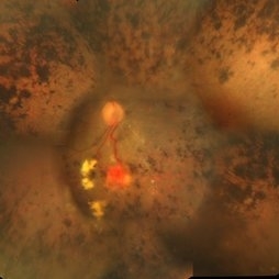 Retinitis Pigmentosa With Hemangioma CF
Dec 15 2016 by Manish Nagpal, MD, FRCS (UK), FASRS
Fluorescein angiography OS of a patient having retinitis pigmentosa with a hemangioma inferiorly.
Condition/keywords: hemangioma, retinitis pigmentosa
-
 Autofluorescence of Retinitis Pigmentosa
Jul 13 2016 by Linda A Cernichiaro- Espinosa, MD
Fundus autofluorescence of an 53-year-old woman with retinitis pigmentosa.
Photographer: Tec Ricardo Montoya, Clínica Oftalmológica Anzures
Condition/keywords: retinitis pigmentosa
-
 Retinitis Pigmentosa
Oct 7 2015 by Avris Romario Diparaja Siahaan
Fundus photograph of a 29-year-old-man with retinitis pigmentosa in both eyes.
Photographer: Yohanes Harry Purwanto, Klinik Mata Nusantara
Imaging device: Topcon TRC 50DX IA
Condition/keywords: color fundus photograph, retinitis pigmentosa
-
 Retinitis Pigmentosa
Apr 14 2014 by Dipankar Barua, M.Sc
Male patient, 18-years-old. On examination his vision is 6/9 both eye. His IOP is 10mmHg in both eye. It seems to be a case of congenital retinitis pigmentosa.
Photographer: Dipankar Barua
Imaging device: Topcon TRC 50 DX (IA)
Condition/keywords: retinitis pigmentosa
-
 Unilateral Retinitis Pigmentosa
May 1 2014 by Raj K. Maturi, MD
53-year-old woman with significant salt and pepper retinopathy OS.
Photographer: Tom Steele, Midwest Eye Institute
Condition/keywords: retinitis pigmentosa (RP) dystrophy
-
 Sector Retinitis Pigmentosa
Mar 13 2014 by Hyung-Woo Kwak, MD
Fundus photograph of an 57-year-old woman with a sector retinitis pigmentosa. Regionalized areas of bone spicule pigmentation is in the inferior quadrants of the retina.
Photographer: Missok Lee, Kyung Hee University Hospital, Seoul, Korea
Imaging device: Zeiss F450 Plus
Condition/keywords: sector retinitis pigmentosa
 Loading…