Search results (5 results)
-
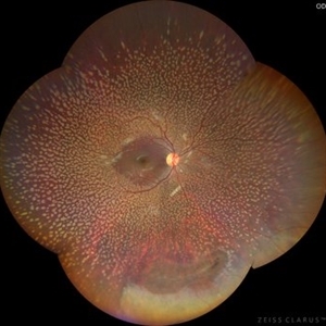 Benign Familial Fleck Retina
Dec 21 2023 by Vishal Agrawal, MD, FRCS,FACS,FASRS
10-year male with high myopia on examination revealed diffuse flecks distributed all over fundus in both eyes sparing macula. Inferior lattice with WWOP areas were also noted in right eye.
Photographer: Dr Ayushi
Imaging device: Clarus 700
Condition/keywords: fleck retinopathy, myopia
-
 Myelinated Nerve Fibre (MNF)
Jun 17 2023 by Harsh Vardhan Singh, MS
Fundus photograph of 32-year-old male having good best corrected visual acuity in both eyes with right eye having high myopia & MNF as incidental finding
Photographer: Dr Harsh Vardhan Singh, Assistant Professor, AIIMS, Guwahati
Condition/keywords: medullated nerve fibers, MNF, myelinated nerve fiber layer, myelinated nerve fibers, Nerve fiber layer arrangements, NFL
-
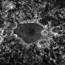 PDR; High Myopia; PRP
May 2 2019 by Carissa Hurdstrom
PDR; high myopia; PRP
Imaging device: Optos
Condition/keywords: fluorescein angiogram (FA), high myopia, pan-retinal photocoagulation (PRP), proliferative diabetic retinopathy (PDR)
-
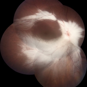
 Submacular Hemorrhage
Mar 12 2016 by Sjakon G Tahija, MD
This a fundus photograph of a high myope who presented with a submacular hemorrhage.
Photographer: Avris Siahaan, Klinik Mata Nusantara, Jakarta, Indonesia
Condition/keywords: high myopia, spontaneous submacular hemorrhage
-
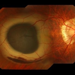
 Foveoschisis secondary to high myopia
Mar 13 2015 by Niloofar Piri, MD
Infrared and HD-OCT of the right eye in a 55-year-old African American female with high myopia (more than -6.00 D), BCVA: 20/25 OU Cartwheel appearance of the fovea in the infrared imaging is visible. HD- OCT demonstartes schisis in different layers of the retina (both NFL and OPL; notice stretching of the Muller cells); VMT is also present . Outer retinal layers are preserved which explains the good vision . She had the same findings in OS.
Photographer: Niloofar Piri, MD
Imaging device: Heidelberg Spectralis
Condition/keywords: high myopia, retinoschisis
 Loading…