Search results (4 results)
-
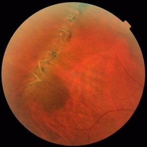
 Choroidal Melanoma
Jan 30 2019 by Karen Panzegrau
Ultra-wide field optos image of a 27-year-old male patient who presented with loss of vision for about 6-8 weeks. Previous choroidal nevus seen. Recommended annual monitoring. No exam for since 10/2014. Brachytherapy vs enucleation was discussed. Brachytherapy was decided as treatment. Full metastatic work up is being performed.
Photographer: Karen Panzegrau
Imaging device: Optos
Condition/keywords: choroidal nevus, exudative retinal detachment, malignant neoplasm of eye, Optos, ultra-wide field imaging
-
 Lattice Degeneration and Choroidal Nevus
Oct 10 2015 by Hamid Ahmadieh, MD
Color fundus photograph of the right eye of a 46-year-old woman with a typical lattice degeneration and an adjacent choroidal nevus.
Photographer: Solmaz Shahmohammad, Negah Eye Center, Tehran, Iran
Condition/keywords: choroidal nevus, color fundus photograph, lattice degeneration
-
 Choroidal Nevus with Subretinal Hemorrhage
Jan 29 2015 by Gordon Finnerty
47-year-old woman diagnosed with choroidal nevus with subretinal hemorrhage.
Photographer: Gordon Finnerty
Imaging device: Topcon TRC -50DX
Condition/keywords: choroidal nevus, subretinal hemorrhage
-
 Choroidal Nevus Myelinated Nerve Fiber Layer
Nov 5 2014 by Raj K. Maturi, MD
85-year-old woman with severe vision loss in left eye. Recommended observation.
Photographer: Tom Steele
Condition/keywords: choroidal nevus, myelinated nerve fiber layer
 Loading…