Search results (11 results)
-
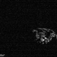 Floating "angle" in Vitreous
Sep 7 2022 by Stephanie Moolman
OCT scan of a vitreous floater in a 42- year-old male after anti-VEGF injection.
Photographer: Stephanie Moolman, Ophthalmic Photographer at Dr Marissa Willemse
Imaging device: Heidelberg Spectralis
Condition/keywords: black floaters, vitreous floaters
-
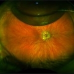
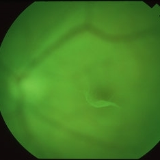 Pre-Macular Floater
Apr 8 2019 by Gary R. Cook, MD, FACS
Red-free photograph of the left eye of a middle-aged white male with a symptomatic pre-macular vitreous condensate/floater seen just ahead of the fovea; V.A. = 20/20
Imaging device: Topcon VT-50
Condition/keywords: floaters, vitreous condensation, vitreous floaters
-
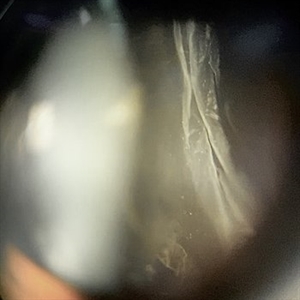 Vitreous Body
Jan 3 2020 by Manuel Ángel Alcántara Delgado, MD
Slit lamp photograph of a 35-year-old woman with history of floaters.
Photographer: Manuel Ángel Alcántara Delgado, CMN SXXI, Mexico City
Condition/keywords: vitreous, vitreous base, vitreous floaters
-
 Vitreous Floater
Dec 18 2017 by Nichole Lewis
Large vitreous floater.
Photographer: Nichole Lewis
Condition/keywords: vitreous floaters
-
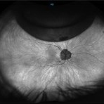
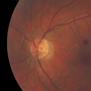 Weiss Ring
Oct 22 2019 by Jessica Norkus
Fundus photo taken on TopCon TRC 50Dx camera of a 60-year-old patient who has experienced an acute PVD. Chief complaint of "large floater" OS prompted exam. Physician noted a significant Weiss Ring and requested fundus color photo for documentation.
Photographer: Jessica Norkus, COA (Retina Specialists of Michigan)
Imaging device: TopCon TRC 50Dx
Condition/keywords: color fundus photograph, color photo, fundus photograph, nerve, optic disc, posterior vitreous detachment, retina, vitreous floaters, Weiss ring
-
 Gas Bubble
May 21 2021 by Raj K. Maturi, MD
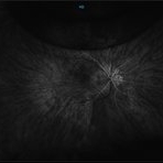
S/P PPV 73-year-old woman with small retinal detachment. Originally presented with decrease vision and residual vitreous floaters. Upon examination small localized retinal detachment with multiple breaks.
Photographer: Charlotte Harris ,Midwest Eye Institute 10300 N Illinois St, Carmel Indiana 46290
Imaging device: Nikon Optos California P200TDx
Condition/keywords: gas bubble
-
 Gas Bubble
May 21 2021 by Raj K. Maturi, MD
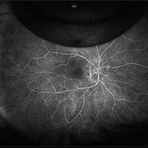
S/P PPV 73-year-old woman with small retinal detachment. Originally presented with decrease vision and residual vitreous floaters. Upon examination small localized retinal detachment with multiple breaks.
Photographer: Charlotte Harris ,Midwest Eye Institute 10300 N Illinois St, Carmel Indiana 46290
Imaging device: Nikon Optos California P200TDx
-
 Gas Bubble
May 21 2021 by Raj K. Maturi, MD
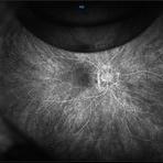
S/P PPV 73-year-old woman with small retinal detachment. Originally presented with decrease vision and residual vitreous floaters. Upon examination small localized retinal detachment with multiple breaks.
Photographer: Charlotte Harris ,Midwest Eye Institute 10300 N Illinois St, Carmel Indiana 46290
Imaging device: Nikon Optos California P200TDx
Condition/keywords: gas bubble
-
 Gas Bubble
May 21 2021 by Raj K. Maturi, MD
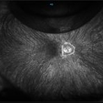
S/P PPV 73-year-old woman with small retinal detachment. Originally presented with decrease vision and residual vitreous floaters. Upon examination small localized retinal detachment with multiple breaks.
Photographer: Charlotte Harris ,Midwest Eye Institute 10300 N Illinois St, Carmel Indiana 46290
Imaging device: Nikon Optos California P200TDx
Condition/keywords: gas bubble
-
 Gas Bubble
May 21 2021 by Raj K. Maturi, MD
S/P PPV 73-year-old woman with small retinal detachment. Originally presented with decrease vision and residual vitreous floaters. Upon examination small localized retinal detachment with multiple breaks.
Photographer: Charlotte Harris ,Midwest Eye Institute 10300 N Illinois St, Carmel Indiana 46290
Imaging device: Nikon Optos California P200TDx
 Loading…