Search results (59 results)
-
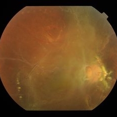
 Age-Related-Maculopathy and Vitreo Macular Adhesion in Left Eye
Feb 7 2018 by Ogugua Ndubuisi Okonkwo, MD, FRCS (Edin), FASRS
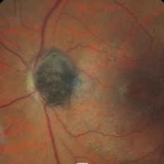
Left eye Fundus photograph of a 73-year-old woman with age related maculopathy , progressive drusenoid deposits and Vitreomacular Adhesion (VMA) monitored over a 10 year duration.
Photographer: Okonkwo Ogugua, Eye Foundation Retina Institute . Lagos
Condition/keywords: drusenoid deposit, vitreomacular traction (VMT)
-
 Age-Related-Maculopathy with Advanced Vitreo Macular Traction in Right Eye
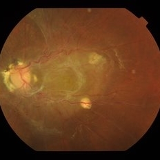
Feb 7 2018 by Ogugua Ndubuisi Okonkwo, MD, FRCS (Edin), FASRS
Right eye Fundus photograph of a 73-year-old woman with age related maculopathy, progressive drusenoid deposits and progressively worsening Vitreomacular Traction (VMT) monitored over a 10 year duration. Requiring surgical release of VMT.
Photographer: Okonkwo Ogugua, Eye Foundation Retina Institute . Lagos
Condition/keywords: drusenoid deposit, vitreomacular adhesion, vitreomacular traction (VMT)
-
 BRVO and VMT Vitreo Macular Traction, Color Photo
Apr 18 2013 by James B. Soque, CRA, OCT-C, COA, FOPS
Color Photograph, 50 degrees, mag 2X of 79-year-old white female, VA sc 20/40, with BRVO OS, and VMT OS, diagnosed on exam and SD OCT. See accompanying RF and FA photos reveal BRVO IT OS, and SD OCT reveals VMT OS.
Photographer: James B. Soque, CRA, COA, Island Retina, Shirley, NY
Imaging device: Topcon TRC-50DX with MERGE software
Condition/keywords: branch retinal vein occlusion (BRVO), vitreomacular traction (VMT)
-
 BRVO and VMT Vitreo Macular Traction, FA Early Phase
Apr 18 2013 by James B. Soque, CRA, OCT-C, COA, FOPS
Early FA photo, 50 degrees, mag 2X of 79-year old white female, VA sc 20/40, with BRVO OS, and VMT OS, diagnosed on exam and SD OCT. See accompanying FC and RF photos reveal BRVO IT OS, and SD OCT reveal VMT OS.
Photographer: James B. Soque, CRA, COA, Island Retina. Shirley, NY
Imaging device: Topcon TRC-50DX with MERGE software
Condition/keywords: branch retinal vein occlusion (BRVO), vitreomacular traction (VMT)
-
 BRVO and VMT Vitreo Macular Traction, Red Free Photo
Apr 18 2013 by James B. Soque, CRA, OCT-C, COA, FOPS
Red Free Photo, 50 degrees, mag 2X of 79-year-old white female, VA sc 20/40, with BRVO OS, and VMT OS, diagnosed on exam and SD OCT. See accompanying FC and FA photos reveal BRVO IT OS, and SD OCT reveal VMT OS.
Photographer: James B. Soque, CRA, COA, Island Retina, Shirley, NY
Imaging device: Topcon TRC-50DX with MERGE software
Condition/keywords: branch retinal vein occlusion (BRVO), vitreomacular traction (VMT)
-
 Combined Hamartoma of Retina and RPE (OD)
Jan 18 2020 by Haider Ali
Fundus photograph of 14-year-old girl with hand movements vision in both eyes. No history of any systemic disease.
Photographer: Haider Ali Chaudhry, Madinah Teaching Hospital, Faisalabad
Condition/keywords: combined hamartoma, epiretinal membrane (ERM), epiretinal membrane formation, vitreomacular traction (VMT)
-
 Combined Hamartoma of Retina and RPE (OS)
Jan 18 2020 by Haider Ali
Fundus photograph of 14-year-old girl with hand movements vision in both eyes. No history of any systemic disease.
Photographer: Haider Ali Chaudhry, Madinah Teaching Hospital, Faisalabad
Condition/keywords: combined hamartoma, epiretinal membrane (ERM), epiretinal membrane formation, neurofibromatosis, vitreomacular traction (VMT)
-
 Eiffel tower within the eye
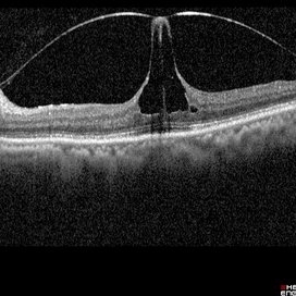
Mar 31 2022 by Bhavani Sankaran, MS (Ophthalmology)
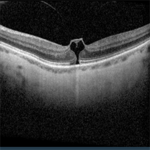
OCT of left eye of a 68 year old female patient who presented with complaints of metamorphopsia. Image depicts vitreomacular traction. BCVA OS 20/40.
Photographer: Dr. Bhavani Sankaran, MS, Aravind Eye Hospital, Madurai
Imaging device: Heidelberg Spectralis
Condition/keywords: vitreomacular interface disorders, vitreomacular traction (VMT)
-
 Enucleated Eye with Retinal Atrophy, Vitreomacular Traction and Keratoconus
May 18 2020 by McGill University Health Centre
This enucleation specimen shows areas of retinal atrophy (*) and areas of vitreomacular traction (arrow). This specimen also demonstrates keratoconus: a degenerative disorder of the eye in which the cornea thins and distorts into a pronounced conical shape (arrowhead). The keratoconus and vitreomacular tractions are unrelated.
Condition/keywords: atrophy, keratoconus, vitreomacular traction (VMT)
-
 Epiretinal Membrane
Sep 26 2018 by Andrea Arriola-Lopez, MD MSc
45-year-old woman, BCVA 20/80 OS. Indirect ophthalmoscopy revealed epiretinal membrane, macular folds and traction. PPV was scheduled.
Photographer: Lourdes Guambo MD, Centro Oftalmológico León, UFM.
Condition/keywords: epiretinal membrane (ERM), vitreomacular interface disorders, vitreomacular traction (VMT)
-
---thumb.jpg/image-square;max$300,300.ImageHandler) ERM and Dry AMD OCT
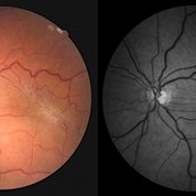
ERM and Dry AMD OCT
Apr 18 2014 by Susanna S. Park, MD, PhD
OCT image of the macula of this 78-year-old man with progressive loss of vision in this left eye from vitreo-macular traction, epiretinal membrane and dry AMD. BCVA 20/100
Photographer: Ellen Redenbo, UC Davis Eye Center
Condition/keywords: dry age-related macular degeneration (dry AMD), epiretinal membrane (ERM), optical coherence tomography (OCT), vitreomacular traction (VMT)
-
 ERMageddon - Wrinkle in the Space-time Fabric of Macula
Oct 29 2025 by SHRADDHA RAJ SHRIVASTAVA
38 year old female with Epiretinal Membrane (ERM) over macula, post laser barrage for multiple symptomatic Horse-shoe Tears (HSTs) and Lattice Degenerations. Posterior pole revealed tilted disc with peripapillary atrophy. There is thick opaque epiretinal membrane obscuring the underlying superior arcade vessels and causing foveal ectopia with distortion of perimacular vasculature. Patient was planned for Right Eye pars plana vitrectomy for ERM peeling.
Photographer: Dr. Shraddha Raj Shrivastava
Imaging device: Nidek Mirante SLO/OCT (Confocal scanning/Spectral domain OCT
Condition/keywords: ectopic fovea, epiretinal membrane (ERM), ERM, horseshoe tear, vitreomacular traction (VMT)
-
 ERMageddon - Wrinkle in the Space-time Fabric of Macula
Oct 29 2025 by SHRADDHA RAJ SHRIVASTAVA
38 year old female with Epiretinal Membrane (ERM) over macula, post laser barrage for multiple symptomatic Horse-shoe Tears (HSTs) and Lattice Degenerations (seen on wide-field image). Posterior pole revealed tilted disc with peripapillary atrophy. There is thick opaque epiretinal membrane obscuring the underlying superior arcade vessels and causing foveal ectopia with distortion of perimacular vasculature. Patient was planned for Right Eye pars plana vitrectomy for ERM peeling.
Photographer: Dr. Shraddha Raj Shrivastava
Imaging device: Nidek Mirante SLO/OCT (Confocal scanning/Spectral domain OCT
Condition/keywords: BARRAGE LASER, ectopic fovea, epiretinal membrane (ERM), horseshoe tear, lattice degeneration, vitreomacular traction (VMT)
-
 Evolution of VMT - OCT 1
Dec 23 2012 by Alex P. Hunyor, MD
OCT on initial presentation, with early separation of posterior cortical vitreous from the temporal perifoveal region.
Condition/keywords: vitreomacular traction (VMT)
-
 Evolution of VMT - OCT 2 at 6 months
Dec 23 2012 by Alex P. Hunyor, MD
OCT scan 6 months later, showing further separation of posterior cortical vitreous with early VMT.
Condition/keywords: vitreomacular traction (VMT)
-
 Evolution of VMT - OCT 3 at 8 months
Dec 23 2012 by Alex P. Hunyor, MD
OCT at 8 months showing further development of focal VMT.
Condition/keywords: vitreomacular traction (VMT)
-
 Evolution of VMT - OCT 4 at 12 months
Dec 23 2012 by Alex P. Hunyor, MD
At 12 months following initial OCT, there has been complete separation of the posterior cortical vitreous from the fovea, with resolution of the VMT.
Condition/keywords: vitreomacular traction (VMT)
-
 Eye Heart You
Jul 23 2024 by Ashley Phillips
VMT causing macular hole.
Photographer: Ashley Phillips
Imaging device: Zeiss 5000
Condition/keywords: vitreomacular traction (VMT)
-
 Macroaneurysm c Ring of HEs and VMT
Jun 15 2016 by Jordan Ball, DO
Fundus photo of 51-year-old male with a temporal macroaneurysm and surrounding ring of exudates and concentric VMT. Pt has 20/30 J2 vision and OD is a prosthesis.
Condition/keywords: macroaneurysm, macular exudates, vitreomacular traction (VMT)
-
 Macular Hole Observed
Dec 2 2017 by John S. King, MD
20/30 initially with some distortion (left); discussed surgery; decided to wait two weeks; progressed some 2 weeks later (right); planned surgery; he was hesitant, so decided to get OCT few days before surgery.
Imaging device: Cirrus
Condition/keywords: macular hole, vitreomacular traction (VMT)
-
 Macular Hole Observed
Dec 2 2017 by John S. King, MD
Few days prior to surgery, hole closed, VMT remains (left); one month later about the same (right).
Imaging device: Cirrus
Condition/keywords: macular hole, vitreomacular traction (VMT)
-
 Macular Hole Observed
Dec 2 2017 by John S. King, MD
Continued to monitor; minimal to no symptoms; about 6 months out, VMT released; vision 20/25+.
Imaging device: Cirrus
Condition/keywords: macular hole, vitreomacular traction (VMT)
-
 Melanocytoma and Vitreomacular Traction Syndrome Fundus Color Image
Mar 19 2018 by Nelson Antonio Segovia Rodríguez, MD
Color fundus image of an 68-year-old male with a optic dis melanocytoma and associated vitreomacular traction syndrome.
Photographer: Nelson Segovia, private practice
Imaging device: Zeiss
Condition/keywords: melanocytoma, optical coherence tomography (OCT), vitreomacular traction (VMT)
-
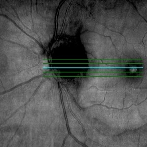 Melanocytoma and Vitreomacular Traction Syndrome OCT Fundus Image
Mar 19 2018 by Nelson Antonio Segovia Rodríguez, MD
OCT fundus image of an 68-year-old male with a optic dis melanocytoma and associated vitreomacular traction syndrome.
Photographer: Nelson Segovia, private practice
Imaging device: Zeiss Cirrus
Condition/keywords: melanocytoma, vitreomacular traction (VMT)
-
 Mild VMT plus drusen
Dec 23 2012 by Alex P. Hunyor, MD
Fundus photograph of the right eye of a 62-year-old female referred with drusen in the right macula. The accompanying OCT scans document the evolution of posterior vitreous separation, with mild vitreomacular traction (VMT) which then spontaneously releases.
Condition/keywords: vitreomacular traction (VMT)
 Loading…