Search results (206 results)
-
25 Gauge Vitrectomy Membrane Shaving
Jan 31 2015 by Thomas A. Ciulla, MD, MBA, FASRS
Membrane shaving of dense membranes in diabetic traction detachment using 25 gauge vitrectomy.
Condition/keywords: diabetes, pars plana vitrectomy (PPV), retina surgery, tractional retinal detachment, vitreoretinal surgery
-
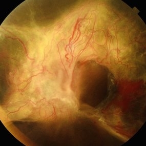
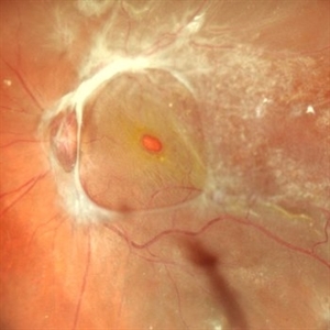
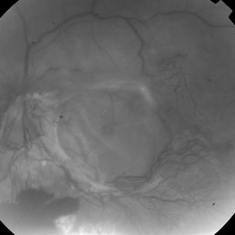
 360 retinotomy for combined closed funnel tractional and rhematogenous retinal detachment
Jan 1 2023 by Malek Yassine, MD
This is Fundus Autofluorecence, showing the residual hypoautofluorescent spots on the exposed choroid, relating to the previous panretinal photocoagulation, as well as the limits of the retinotomy with continuous laser which appeasr hypoautofluorecent with hyperautofluorecent margins.
Photographer: Malek Yassine, HMOED, Agadir, Morocco.
Imaging device: Zeiss Clarus
Condition/keywords: combined retinal detachment, rhegmatogenous retinal detachment, tractional retinal detachment
-
 Active Proliferative Diabetic Retinopathy
Aug 16 2022 by Donnie Willis
51 yo female. Uncontrolled Diabetes. Active Proliferative Diabetic Retinopathy.
Photographer: Donnie Willis, Tennessee Retina
Imaging device: Optos
Condition/keywords: capillary dropouts, Diabetes, fluorescein angiogram (FA), OPTOS, proliferative diabetic retinopathy (PDR), tractional retinal detachment
-
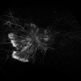
 Acute Posterior Vitreous Detachment
Nov 9 2012 by Norman Byer
This large and complicated retinal tear in a 51-year-old man resulted from an acute posterior vitreous detachment which concentrated its tractional forces around this area of lattice degeneration. Because of the powerful traction, there is an additional central tear splitting the large retinal flap and almost severing one of its arms. The traction was strong enough to completely rupture the blood vessel just to the left of the flap. Marking the ruptured peripheral end of the blood vessel is a yellow depigmented thrombus.
Condition/keywords: acute posterior vitreous detachment, depigmented thrombus, lattice degeneration, retinal tear, tractional retinal detachment
-
 Acute Retinal Necrosis with Proliferative Vitreoretinopathy and Total Retinal Detachment
Mar 26 2019 by Gary R. Cook, MD, FACS
Same WF patient 9 weeks after initial presentation with Acute Retinal Necrosis now with proliferative vitreoretinopathy and a total combined traction & rhegmatogenous retinal detachment
Imaging device: Topcon VT-50
Condition/keywords: acute retinal necrosis, proliferative vitreoretinopathy (PVR), tractional retinal detachment
-
 Advanced PDR
Sep 29 2012 by Hamid Ahmadieh, MD
Color fundus photograph of a 32-year-old man with insulin-dependent diabetes mellitus and regressed advanced PDR with severe fibrous proliferation and traction retinal detachment sparing the macula.
Photographer: Hamid Ahmadieh, MD; Ophthalmic Research Center, Labbafinejad Medical Center, Shahid Beheshti University of Medical Sciences
Condition/keywords: fibrous proliferation, tractional retinal detachment
-
 Advanced Proliferative Diabetic Retinopathy
Apr 9 2025 by Gustavo Uriel Fonseca Aguirre
B-mode ultrasound of a patient with long-standing poorly controlled diabetes demonstrates characteristic findings of advanced proliferative diabetic retinopathy. The examination reveals moderate vitreous hemorrhage appearing as diffuse hyperechoic opacities throughout the vitreous cavity, along with a posterior hyaloid membrane densely infiltrated by hemorrhagic material, showing irregular thickening and increased reflectivity. A mild subhyaloid hemorrhage is visible as a subtle hyphema-like space anterior to the retinal surface. The study documents a total tractional retinal detachment, evidenced by rigid retinal folds with clear insertion points of vitreous strands, accompanied by a significant subretinal hemorrhage seen as a prominent hyperechoic collection beneath the elevated retina. These findings collectively illustrate the severe vitreoretinal interface pathology characteristic of end-stage diabetic eye disease, with predominant tractional components and distinct echographic stratification of hemorrhagic layers - from anterior vitreous involvement to deeper subretinal blood accumulation.
Photographer: Gustavo U. Fonseca Aguirre, Hospital Conde de Valenciana, Ciudad de México
Condition/keywords: diabetic retinopathy, tractional retinal detachment, Vitreous hemorrhage
-
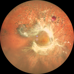
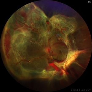
 Annular Tractional Retinal Detachment
Jul 5 2025 by César Adrián Gómez Valdivia, MD
Fundus photograph of an 66 YO female patient diagnosed with advanced proliferative diabetic retinopathy.
Photographer: @eyemissu2
Imaging device: TOPCON TRC-50DX
Condition/keywords: tractional retinal detachment
-
 Annular Tractional Retinal Detachment
Jul 4 2024 by Hector Gabriel Moreno Solano, MD, MHA
52-year-old Hispanic female patient with a diagnosis of type II diabetes mellitus of 15 years of evolution, comes to the retina service for progressive visual loss in the right eye (single functional eye) with visual acuity of 20/100, Fundus examination reveals laser-modified proliferative diabetic retinopathy with activity + annular tractional retinal detachment with macular involvement.
Photographer: Hector Gabriel Moreno Solano, MD, MHA, HGZ #20 IMSS Puebla.
Imaging device: Mirante
Condition/keywords: macular detachment, proliferative diabetic retinopathy (PDR), tractional retinal detachment
-
000---thumb.jpg/image-square;max$300,300.ImageHandler) Anterior Segment Photo of Emulsified Silicone Oil
Anterior Segment Photo of Emulsified Silicone Oil
Dec 25 2013 by Dong Yoon Kim, MD
47-year-old woman underwent vitrectomy and silicone oil tampoande for tractional retinal detachment due to proliferative diabetic retinopathy. 8 months after silicone oil tamponade, silicone oil was emulsified. And emulsified silicone oil was observed at anterior chamber.
Condition/keywords: silicone oil, tractional retinal detachment
-
 Before and After Vitrectomy
Nov 17 2023 by Bradley T. Smith, MD, FASRS
Middle age male diabetic retinopathy and resolving exudate following repair of tractional detachment with membrane peeling.
Condition/keywords: coats-like response, Diabetes, fibrotic neovascularization, fibrovascular proliferation, pars plana vitrectomy (PPV), proliferative diabetic retinopathy (PDR), tractional retinal detachment
-
 Combined Pathology
Oct 26 2024 by rahul saradge
53 year old male patient was presented with a complaints of diminished vision in LE since 1 month. The BCVA in RE was 6/36p and LE was CF 1/2m. Ocular dilated examination showed RE temporal CD with ?CRVO,OIS and OS showed TRD and old Hemi CRVO. Patient was injected with PST tricot followed by PRP laser at an interval of 1 week. Patient improved to BCVA 6/9.
Photographer: Aishwarya Bangar Isha Netralaya Thane
Imaging device: optos
Condition/keywords: choroidal detachment, crvo, ois, optos, pan retinal photocoagulation, tractional retinal detachment
-
 Combined Retinal Detachment With Macular Hole
Sep 28 2024 by Tejaswita Verma
Fundus image of the LE of a 67 year old diabetic, hypertensive female with CF 3metres vision showing combined RD with FTMH, in a pseudophakic eye. She was lost to follow up status post 2 anti VEGF injections received 8 months back due to typhoid fever.
Photographer: DR. TEJASWITA VERMA
Imaging device: MIRANTE
Condition/keywords: full thickness macular hole, proliferative diabetic retinopathy (PDR), tractional retinal detachment
-
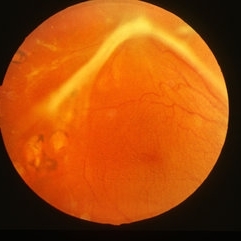
 Combined Tractional and Rhegmatogenous Retinal Detachment
Jan 30 2023 by Olivia Rainey
Ultra-widefield fluorescein angiography of a combined tractional and rhegmatogenous retinal detachment repair affecting the left eye. The retina is attached following silicone oil placement during most recent surgery. The patient was seeing CF at the time the image was taken.
Photographer: Olivia Rainey, OCT-C, COA
Imaging device: Optos California
Condition/keywords: diabetes, diabetic macular edema, diabetic retinopathy, fluorescein angiogram (FA), hyperfluorescence, right eye, scleral buckle, silicone oil, tractional retinal detachment, ultra-wide field imaging, ultra-widefield image
-
---thumb.jpg/image-square;max$300,300.ImageHandler) Complications of ARN, TRD
Complications of ARN, TRD
Feb 27 2013 by Henry J. Kaplan, MD
Development of TRD as a late complication of ARN.
Condition/keywords: acute retinal necrosis, ARN complications, tractional retinal detachment
-
 Diabetes Proliferative
Jul 11 2013 by Jerald A. Bovino, MD
No history, traction retinal detachment, part of stereo pair.
Condition/keywords: diabetic mellitus, stereo pair, tractional retinal detachment
-
 Diabetic Macular TRD
Jan 10 2020 by Somnath Chakraborty, MD
Fundus Montage image of the left eye of a 48-year-old type 2 diabetic with post PRP macular extensive tractional retinal detachment involving macula.
Photographer: Pulak Roy
Condition/keywords: diabetic retinopathy, proliferative diabetic retinopathy (PDR), tractional retinal detachment, vitrectomy, vitreomacular surgery
-
 Diabetic Retinopathy
Dec 11 2019 by Lauren Whaley
44-year-old male diabetic patient had an acute change in A1C over 9 months and ended up with a tractional retinal detachmen in right eye. This photo is 2 weeks post operative with current vision level at hand motion. He had extensive laser, retinectomy, and silicone oil fill.
Photographer: Lauren R. Whaley, COA
Imaging device: Optos Wide Field
Condition/keywords: diabetes, diabetic retinopathy, fibrosis, laser scarring, proliferative vitreoretinopathy (PVR), retinectomy, silicone oil, tractional retinal detachment
-
 Diabetic traction retinal detachment
Jan 9 2023 by JORGE SOBERANES
Proliferative diabetic retinopathy with extensive traction retinal detachment in a patient with type 1 diabetes mellitus.
Photographer: Dr. Jorge I. Soberanes, Asociación para Evitar la Ceguera en México.
Imaging device: Zeiss Clarus 700
Condition/keywords: Retinal Detachment, tractional retinal detachment
-
 Diabetic Tractional Retinal Detachment
Jan 10 2018 by Peter H. Tang, MD, PhD
Fundus photograph of a 46-year-old woman with proliferative diabetic retinopathy and tractional retinal detachment that is poorly controlled.
Imaging device: Optos California
Condition/keywords: diabetes, neovascularization elsewhere (NVE), neovascularization of the disc (NVD), pan-retinal photocoagulation (PRP), proliferative diabetic retinopathy (PDR), retinal fibrosis, tractional retinal detachment
-
 Diabetic TRD
Jun 20 2016 by John S. King, MD
Poorly controlled DM missed many appointments due to CVA came in with complaint of floaters; initial red free; CF.
Condition/keywords: diabetic traction detachment, tractional retinal detachment
-
 Diabetic TRD
Jun 20 2016 by John S. King, MD
3 and 6 m post-op 2 avastins 20/300 at 6 months c 3+NSC
Condition/keywords: diabetic traction detachment, tractional retinal detachment
-
 Diabetic TRD
Jun 20 2016 by John S. King, MD
Poorly controlled DM missed many appointments due to CVA came in with complaint of floaters; initial red free; CF initial visit and three months post-op.
Condition/keywords: diabetic traction detachment, tractional retinal detachment
 Loading…