Search results (22 results)
-
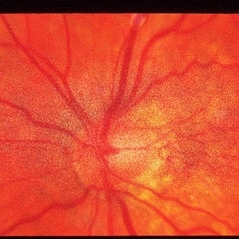
 Disc Hypoplasia
Feb 20 2013 by From the Collections of Thomas M. Aaberg, MD and Thomas M. Aaberg Jr., MD
Disc hypoplasia; tilted disc and field defect.
Condition/keywords: disc hypoplasia, tilted disc
-
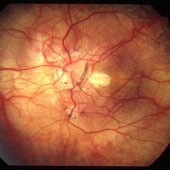
 Disc, Tilted
Apr 4 2014 by H. Michael Lambert, MD
Tilted Disc with peripapillary atrophy
Condition/keywords: tilted disc
-
 Dislocated Intraocular Lens, Tilted Disc, Posterior Staphyloma.
Aug 18 2021 by Jesus Lozano, MD
Fundus photograph of 80-year-old woman, single eye with left eye posterior dislocation of lens, tilted disc and posterior staphyloma.
Photographer: Yair Bet Yosef, Hadassah Medical Center. Israel
Imaging device: Optos Silverstone
Condition/keywords: posterior dislocation of lens, posterior staphyloma, tilted disc
-
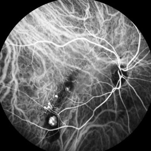
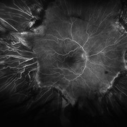 Fluorescein Angiogram of ROP With Cryo Scarring
Jul 7 2025 by Jenn Geelan
FA photo of a 34 year old male with prior stage 3 ROP with history of 360 degree cryotherapy.
Photographer: Jenn Geelan, Retina-Vitreous Surgeons of CNY
Imaging device: Optos California
Condition/keywords: cryotheraphy scar, fluorescein angiogram (FA), fundus photograph, retinopathy of prematurity (ROP), ROP, tilted disc
-
 High Myopia
May 2 2013 by Henry J. Kaplan, MD
Chorioretinal atrophy in high myopia and tilted disc.
Condition/keywords: high myopia, tilted disc
-
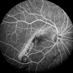
 Myopic Maculoscheisis
Sep 11 2013 by Jason S. Calhoun
Fluorescence angiography and fundus photography shows RPE mottling and staphyloma in the right eye. VA is 20/50, right eye. No subretinal fluid found.
Photographer: Jason S. Calhoun, Department of Ophthalmology, Mayo Clinic Jacksonville, Florida
Imaging device: TOPCON TRC 50-EX
Condition/keywords: myopia, tilted disc
-
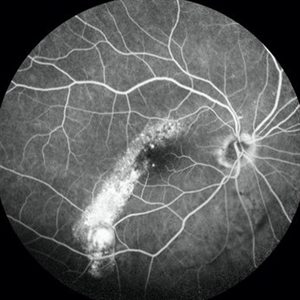
 Tilted Disc Syndrome Complicated with RPE Atrophy and Polypoidal Choroidal Vasculopathy
Jan 20 2020 by Pierre-Henry Gabrielle, MD
Early-phase FA angiography of an 81-year-old woman with a tilted disc syndrome complicated with RPE atrophy and polypoidal choroidal vasculopathy.
Photographer: Pierre-Henry Gabrielle, Ophthalmology department, Dijon University Hospital, France.
Imaging device: Heidelberg spectralis angiography
Condition/keywords: atrophic pigment epithelium, fluorescein angiogram (FA), polypoidal choroidal vasculopathy (PCV), tilted disc
-
 Tilted Disc Syndrome Complicated with RPE Atrophy and Polypoidal Choroidal Vasculopathy
Jan 20 2020 by Pierre-Henry Gabrielle, MD
Early-phase ICG angiography of an 81-year-old woman with a tilted disc syndrome complicated with RPE atrophy and polypoidal choroidal vasculopathy.
Photographer: Pierre-Henry Gabrielle, Ophthalmology department, Dijon University Hospital, France.
Imaging device: Heidelberg spectralis angiography
Condition/keywords: atrophic pigment epithelium, indocyanine green (ICG) angiography, polypoidal choroidal vasculopathy (PCV), tilted disc
-
 Tilted Disc Syndrome Complicated with RPE Atrophy and Polypoidal Choroidal Vasculopathy
Jan 20 2020 by Pierre-Henry Gabrielle, MD
Fundus optos photograph of an 81-year-old woman with a tilted disc syndrome complicated with RPE atrophy and polypoidal choroidal vasculopathy.
Photographer: Pierre-Henry Gabrielle, Ophthalmology department, Dijon University Hospital, France.
Imaging device: Optos
Condition/keywords: atrophic pigment epithelium, indocyanine green (ICG) angiography, polypoidal choroidal vasculopathy (PCV), tilted disc
-
 Tilted Disc Syndrome Complicated with RPE Atrophy and Polypoidal Choroidal Vasculopathy
Jan 20 2020 by Pierre-Henry Gabrielle, MD
Late-phase fluorescein angiography of an 81-year-old woman with a tilted disc syndrome complicated with RPE atrophy and polypoidal choroidal vasculopathy.
Photographer: Pierre-Henry Gabrielle, Ophthalmology department, Dijon University Hospital, France.
Imaging device: Heidelberg spectralis angiography
Condition/keywords: atrophic pigment epithelium, fluorescein angiogram (FA), polypoidal choroidal vasculopathy (PCV), tilted disc
-
 Tilted Disc Syndrome Complicated with RPE Atrophy and Polypoidal Choroidal Vasculopathy
Jan 20 2020 by Pierre-Henry Gabrielle, MD
Late-phase ICG angiography of an 81-year-old woman with a tilted disc syndrome complicated with RPE atrophy and polypoidal choroidal vasculopathy.
Photographer: Pierre-Henry Gabrielle, Ophthalmology department, Dijon University Hospital, France.
Imaging device: Heidelberg spectralis angiography
Condition/keywords: atrophic pigment epithelium, indocyanine green (ICG) angiography, polypoidal choroidal vasculopathy (PCV), tilted disc
-
 Tilted Disc Syndrome Complicated with RPE Atrophy and Polypoidal Choroidal Vasculopathy
Jan 20 2020 by Pierre-Henry Gabrielle, MD
Coupled OCT B-scan and ICG angiography of an 81-year-old woman with a tilted disc syndrome complicated with RPE atrophy and polypoidal choroidal vasculopathy.
Photographer: Pierre-Henry Gabrielle, Ophthalmology department, Dijon University Hospital, France.
Imaging device: Heidelberg spectralis
Condition/keywords: atrophic pigment epithelium, indocyanine green (ICG) angiography, optical coherence tomography (OCT), polypoidal choroidal vasculopathy (PCV), tilted disc
-
 Tilted Disc Syndrome Complicated with RPE Atrophy and Polypoidal Choroidal Vasculopathy
Jan 20 2020 by Pierre-Henry Gabrielle, MD
Zoomed fundus optos photograph of an 81-year-old woman with a tilted disc syndrome complicated with RPE atrophy and polypoidal choroidal vasculopathy.
Photographer: Pierre-Henry Gabrielle, Ophthalmology department, Dijon University Hospital, France.
Imaging device: Optos
Condition/keywords: atrophic pigment epithelium, Optos, polypoidal choroidal vasculopathy (PCV), tilted disc
-
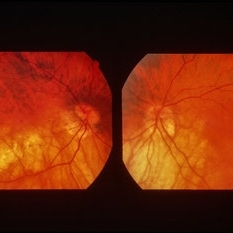
 Inferior Choroidal Coloboma and Tilted Disc
Feb 19 2013 by From the Collections of Thomas M. Aaberg, MD and Thomas M. Aaberg Jr., MD
NLP; Left of stereo pair.
Condition/keywords: coloboma, stereo pair
-
 Inferior Choroidal Coloboma and Tilted Disc
Feb 19 2013 by From the Collections of Thomas M. Aaberg, MD and Thomas M. Aaberg Jr., MD
NLP; Right of stereo pair.
Condition/keywords: coloboma, stereo pair
-
 Inferior Choroidal Coloboma and Tilted Disc
Feb 19 2013 by From the Collections of Thomas M. Aaberg, MD and Thomas M. Aaberg Jr., MD
Inferior choroidal coloboma and tilted disc.
Condition/keywords: coloboma
-
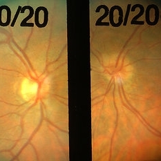
 Myopia
Apr 24 2014 by Howard Schatz, MD
80-year-old white female. Myopia (tilted disc). 20/50 OU.
Condition/keywords: myopia
-
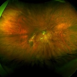
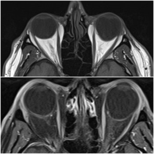 Staphyloma in Pathologic Myopia
Feb 7 2020 by Jonathan C. Tsui, MD
A 51-year-old presents with a six-month history of OS vision loss found to be CF at 3'. Fundus exam demonstrated pathologic myopia OS>OD with tilted discs. MRI Orbit Axial T1 images demonstrate significant compatible findings of staphylomas OS>OD.
Condition/keywords: pathologic myopia, staphyloma
-
 Unilateral Tilted Disc
Feb 20 2013 by From the Collections of Thomas M. Aaberg, MD and Thomas M. Aaberg Jr., MD
30-year-old.
Condition/keywords: unilateral tilted disc
 Loading…