Search results (9 results)
-
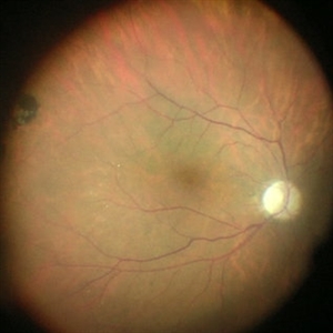
 Giant Retinal Tear After Buckle With Perfluorocarbon Liquid
Dec 24 2013 by Gregg T. Kokame, MD, MMM, FASRS
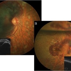
Well-centered IOL after repair of giant retinal tear post buckle with perfluorocarbon liquid. Patient had history of blunt trauma to the eye and underwent scleral fixation of IOL five years prior, presented with retinal detachment with a giant retinal tear.
Condition/keywords: blunt trauma, surgical management
-
 ILM staining
Dec 11 2019 by Jennifer R Gallagher, MD
Intra-operative funds photo of the macula after ICG staining with removal of excess dye from the vitreous cavity.
Photographer: Hamzah Khalaf, UT Health San Antonio, University Hospital
Condition/keywords: internal limiting membrane (ILM) peeling, staining, surgical management
-
 ILM Staining
Dec 11 2019 by Jennifer R Gallagher, MD
Intra-operative photo of the injection of indocyanine green (ICG) to stain the internal limiting membrane (ILM).
Photographer: Hamzah Khalaf, UT Health San Antonio, University Hospital
Condition/keywords: ILM staining, internal limiting membrane (ILM) peeling, surgical management
-
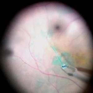 ILM visibility with ICG
Dec 11 2019 by Jennifer R Gallagher, MD
Intra-operative photo highlighting the utility of ICG for ILM visibility.
Photographer: Hamzah Khalaf, UT Health San Antonio, University Hospital
Condition/keywords: internal limiting membrane (ILM) peeling, staining, surgical management
-
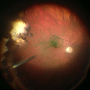
 Scleral-Buckle in Rhegmatogenous Retinal Detachment
Dec 18 2019 by VERONICA ADRIANA ROMERO- MORALES, MD
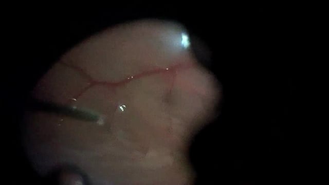
Trans-surgical photograph of a 40-year-old man with phakic eye, high myopia and macula off rhegmatogenous retinal detachment. Without complications after 5 years of follow up.
Photographer: Amanda Estrada López MD, GOVisión, Queretaro, Mexico.
Imaging device: Smartphone
Condition/keywords: high myopia, scleral buckle, surgical management
-
 Submacular Hemorrhage
Jun 14 2018 by Deepak Bhojwani, MS
A 67-year-old lady presented with 10 days old submacular hemorrhage with poor visual acuity of hand movements only(A). She underwent successful surgical pneumatic displacement & ANTI-VEGF injection with good visual outcome(B) gaining 20 letters on ETDRS vision charts.
Photographer: Dr Deepak Bhojwani, Raghudeep Eye Hospital, Ahmedabad
Imaging device: Zeiss Visucam 500
Condition/keywords: submacular hemorrhage, surgical management
-
 Surgical Management of a Symptomatic Full Thickness Macular Fold
Jul 19 2021 by Anton Orlin, MD
A balanced salt solution is injected into the subretinal space to focally detach the macula, ensuring that the fold is incorporated, in order to stretch and relax it. This can be done with a 38 or 41 gauge subretinal cannula, and is typically performed just within the macular arcade. Perfluorocarbon heavy liquid (PFCL) is then injected to flatten the macula and push the excess fluid to the periphery. A peripheral retinotomy is made, and the subretinal fluid is subsequently drained with air fluid exchange. The PFCL is removed and endolaser is applied to surround the retinotomy site. The eye was left with a gas tamponade.
Condition/keywords: macular fold, surgical management, video
-
 Surgical Management of Trauma
Dec 10 2012 by Yale L. Fisher, MD
Dr. Steve Charles discusses his primary steps in managing ocular trauma.
Condition/keywords: surgical management
-
 Surgical Management of Massive Suprachoroidal Hemorrhage: Don’t Play It Blind!
Apr 6 2018 by Jay Sheth
Expulsive suprachoroidal hemorrhage(SCH) is a catastrophic complication of intraocular surgery. Current management includes SCH drainage through external sclerotomies & intermittent fundus evaluation by IO. We describe a novel surgical technique, utilizing chandelier-assisted wide-angled visualization of various steps of SCH drainage in 62/M. Using the wide-angle viewing system & 23G extrusion cannula through asclerotomy, active drainage of SCH was performed whereby we beautifully demonstrate separation of the kissing choroids with gradual unmasking of macula &disc underneath. Post-operatively, patient improved to CF3m & eye was successfully salvaged. Our educational video demonstrates that chandelier-assisted controlled drainage of SCH under continuous visualization is an easy technique to achieve excellent anatomical & visual outcomes with better safety profile. It can be instrumental in training residents & fellows who can simultaneously visualize surgical steps along with surgeon.
Condition/keywords: suprachoroidal hemorrhage, video
 Loading…